
A 47-year-old with a 2 cm lesion in the left hepatic lobe needs nuclear confirmation of a hemangioma before the oncology team sets the follow-up plan. The unit has SPECT/CT, technetium-99m pertechnetate, and in vitro red-cell labeling supplies. Which study plan best demonstrates slow blood pooling in the lesion?
Select an answer to reveal the explanation.
Short Explanation
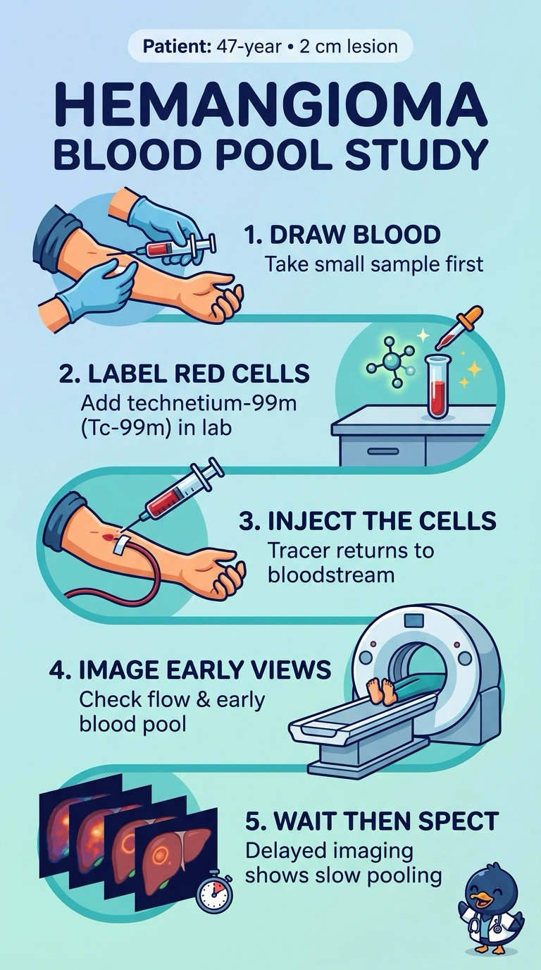
A hemangioma fills slowly, so you need tracer that stays in the bloodstream long enough to pool there and show up again later. Labeling the patient's own red cells keeps activity predominantly in the blood pool for the early and delayed views. It's like watching a sponge fill at its own pace with blood-pool imaging rather than one quick snapshot.
Full Explanation
In vitro technetium-99m red-cell labeling keeps the tracer within the circulating blood pool, allowing a hemangioma’s slow filling and persistent blood-pool activity to be assessed. Flow and early blood-pool images establish the initial vascular pattern, while delayed SPECT localizes retained activity within the lesion and provides tomographic confirmation. In vivo red-cell labeling is a recognized blood-pool technique and could demonstrate pooling, but it disregards the scenario’s specified in vitro labeling supplies and protocol. Delayed planar views can show persistent activity, but they do not meet the stated need for SPECT localization of this small hepatic lesion. Omitting flow images leaves out the initial perfusion phase, so the plan does not provide the requested flow-to-pooling sequence even though early and delayed blood-pool imaging are included. Exam caveat: persistent red-cell activity supports hemangioma but should be interpreted with the imaging pattern and anatomic findings, rather than treated as diagnostic in isolation. Practical check: confirm adequate labeling and review the acquisition timing and SPECT/CT alignment with the lesion’s location.