
The day before a sentinel node biopsy for melanoma on the patient's left forearm, the surgeon schedules preoperative lymphoscintigraphy with Tc-99m filtered sulfur colloid. The tracer must enter lymphatic channels beside the lesion, reach the first draining node, and be imaged while it is still moving. Which injection plan best delivers tracer to the first draining node?
Select an answer to reveal the explanation.
Short Explanation
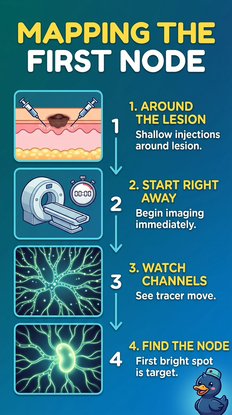
You get the best map by placing several small injections intradermally around the lesion and starting imaging right away, while the tracer is moving toward the first draining node. The old excision scar is not the site described, nanocolloid is not the specified tracer, and waiting an hour can miss the initial drainage sequence.
Full Explanation
Several small intradermal injections around the melanoma place the specified Tc-99m filtered sulfur colloid in superficial lymphatic-rich tissue beside the lesion. Starting imaging right away lets you follow tracer transit and identify the first draining node while the tracer is moving. Injecting around an old excision scar can be useful when the primary lesion has already been removed, but this case describes an existing forearm lesion, so the injections should be placed around that lesion. Tc-99m nanocolloid is used for sentinel-node mapping in some settings, but it does not satisfy the stated requirement to use filtered sulfur colloid. Waiting an hour before imaging may still reveal a node, but it misses the requested moving-tracer phase and may obscure the initial drainage sequence. Exam caveat: mapping protocols and accepted radiocolloids vary by institution, so a generally valid technique may still fail a question’s explicit tracer, injection-site, or timing constraints. Practical check: confirm the injection sites, radiopharmaceutical, and acquisition start time against the lesion location and the local nuclear-medicine protocol.