
A small well-circumscribed lesion is seen in the right lobe of the liver on ultrasound, and the radiologist wants to know whether it is a hemangioma. The patient is booked for in-vitro labeled red blood cell imaging, and SPECT is available in the department. Which acquisition plan will show the lesion's blood pool behavior most reliably?
Select an answer to reveal the explanation.
Short Explanation
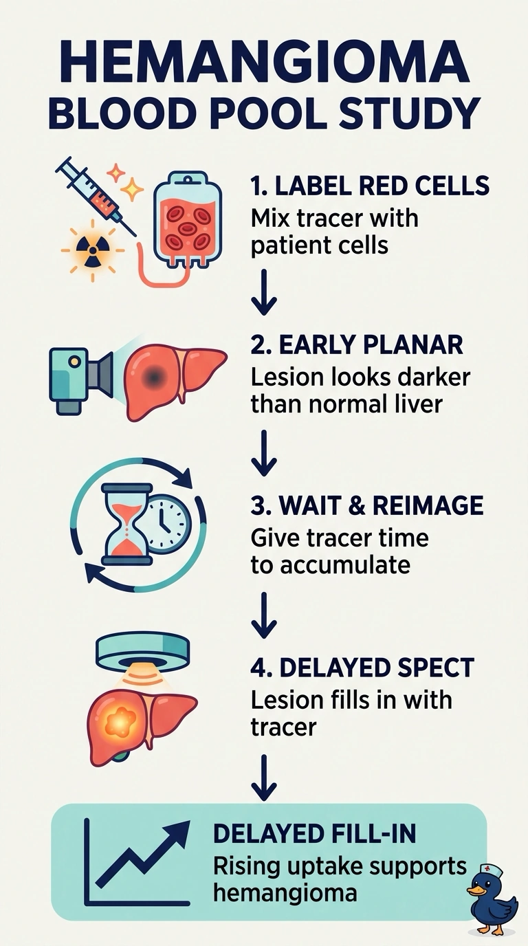
A hemangioma is a slow-filling blood space, so a single early picture can make it look like a cold hole. Pairing early planar imaging with delayed SPECT lets you see the tracer accumulate, which is a characteristic finding. It works like a sponge soaking up water, where the change over time tells the story.
Full Explanation
Hepatic hemangiomas are vascular spaces filled with slowly moving blood, so labeled red cells may accumulate there gradually while normal liver tissue shows activity early in the study. Imaging early and again after a delay lets you see a photopenic area fill in toward or above surrounding liver; delayed SPECT can help localize activity by reducing the effects of overlying structures. Stopping after early planar images misses delayed fill-in, and ending the study after flow images leaves no later blood-pool comparison. Sulfur colloid evaluates Kupffer cell function rather than the vascular filling assessed by labeled red cells. Not every hemangioma fills completely, so a negative delayed study does not exclude the diagnosis and may require correlation with other imaging.