
A patient with a small, rounded lesion in the right lobe of the liver on ultrasound is sent for a nuclear medicine study, and the radiologist suspects a hemangioma and wants images showing it fill with blood over time. The exam must be finished in a single visit and the lesion is only about two centimeters wide. Which liver blood-pool study plan meets all of these requirements?
Select an answer to reveal the explanation.
Short Explanation
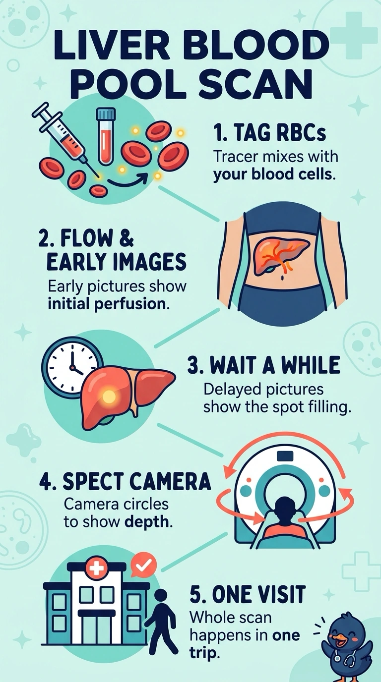
You need a study that follows blood itself, since a hemangioma is a tangle of slow-filling vascular spaces. Labeled red blood cells stay in the bloodstream, so the lesion looks pale at first and fills on delayed images. Flow, early blood-pool, and delayed SPECT together meet your requirements.
Full Explanation
The clinical question is whether a focal liver lesion is a vascular space rather than a solid tumor, and labeled red blood cells answer it because they remain in the vascular compartment long enough to show progressive fill-in. Flow images show initial perfusion; blood-pool and delayed images assess the lesion’s characteristic progressive accumulation. Early blood-pool images allow comparison with surrounding liver, while delayed SPECT captures slow accumulation in cross-section, important for a lesion only about two centimeters wide. Sulfur colloid maps reticuloendothelial phagocytosis instead: it may show a photon-deficient area but cannot demonstrate whether that area fills with blood over time. A hepatobiliary tracer follows liver function and excretion through the liver and gallbladder, addressing obstruction or contractility rather than the vascular nature of a mass. Planar blood-pool images alone can show fill-in, but without tomographic data, a small lesion within liver activity may be missed or mistaken for background. Exam caveat: some departments add computed tomography for attenuation correction, but blood-pool timing still drives the diagnosis. Practical check: confirm labeling efficiency and the delayed time point before starting, because poor labeling or an early final set can mimic a photopenic lesion.