
A 54-year-old outpatient has a 2 cm hypervascular lesion in the right hepatic lobe found incidentally on an abdominal CT after a minor traffic collision. The surgeon wants noninvasive confirmation that the lesion is a hemangioma before considering a biopsy, and the patient can return for a later appointment. Which radiopharmaceutical study and imaging plan should be scheduled to characterize the lesion as a hemangioma?
Select an answer to reveal the explanation.
Short Explanation
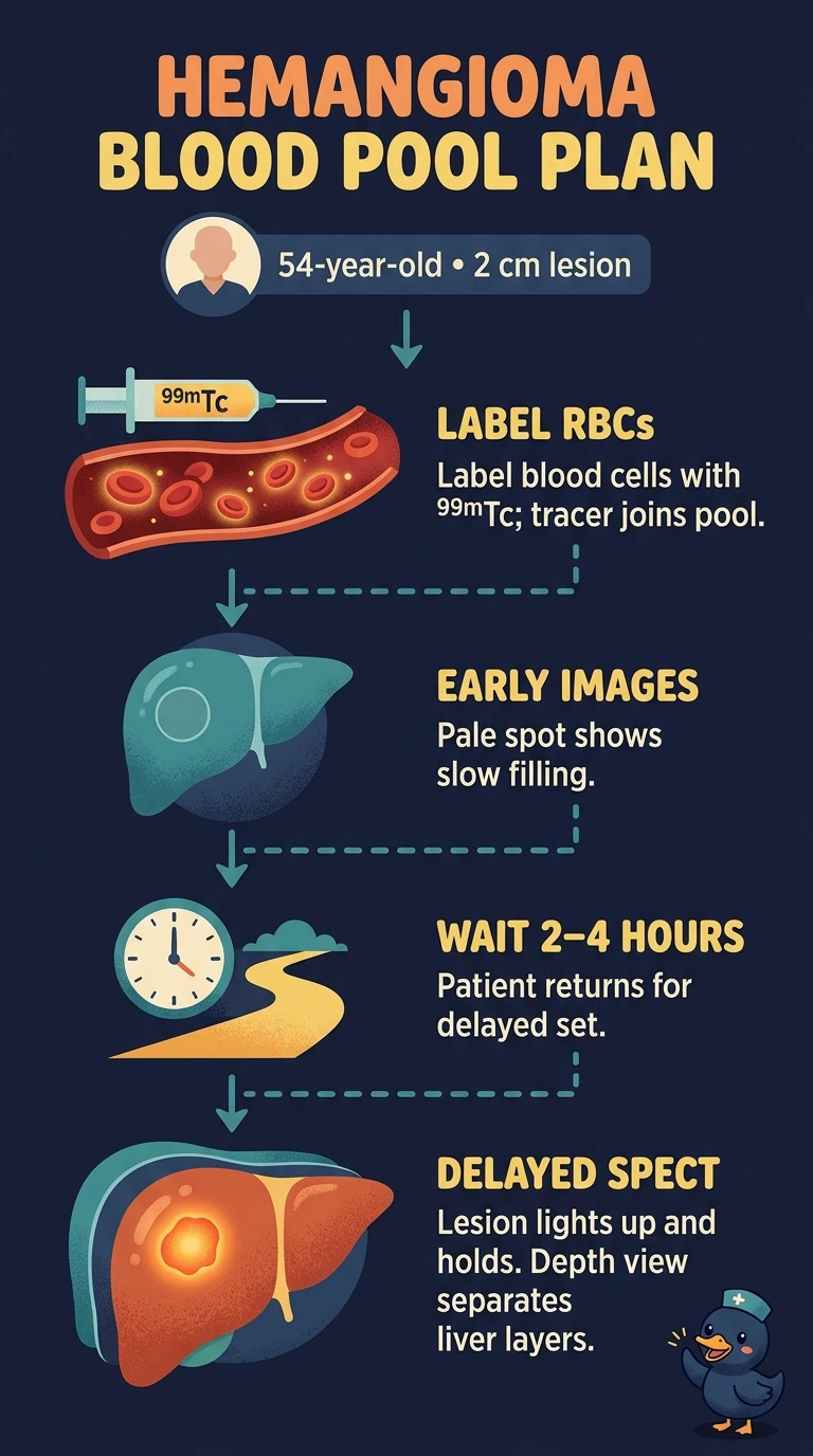
You can characterize a hemangioma with tagged red cells because they remain in the blood pool rather than entering the bile ducts. Early blood-pool images followed by delayed SPECT show the lesion’s gradual filling, and SPECT adds depth information beyond planar images.
Full Explanation
Technetium-99m labeled red blood cells circulate with your own blood. A hemangioma is a slow-flow vascular space that fills late and retains the tagged cells, so it appears photon-poor on early blood-pool images and brighter than the surrounding liver on delayed images; SPECT helps separate overlapping structures and improves depth localization. Technetium-99m sulfur colloid maps Kupffer cell uptake, so a focal photopenic defect does not establish that a lesion is vascular or confirm hemangioma. Technetium-99m mebrofenin follows hepatocyte handling and biliary excretion, which address hepatobiliary function rather than blood-pool retention. The gastrointestinal bleeding study tracks free tracer escaping into the bowel lumen, not a focal hepatic mass. Exam caveat: hemangioma protocols may use in vivo, in vitro, or combined red-cell labeling; the labeling approach changes preparation time, not the imaging goal. Practical check: confirm that delayed blood-pool imaging, including SPECT at 2–4 hours, is scheduled before you leave.