
A rest and stress myocardial perfusion SPECT study on a 58-year-old with a low-risk stress test shows a reversible defect along the inferior wall on the uncorrected images, and the hybrid scanner can also acquire CT. Which processing approach will best show whether soft-tissue attenuation explains the finding?
Select an answer to reveal the explanation.
Short Explanation
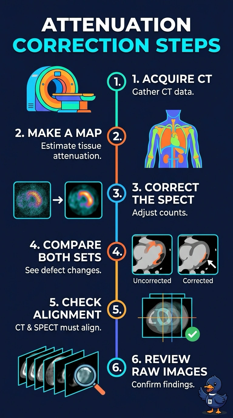
You’ll use CT-based attenuation correction, then compare the corrected and uncorrected perfusion images to see whether the inferior-wall defect fades. The CT map accounts for tissue blocking photons—like adjusting a photo for uneven lighting—and helps show whether attenuation correction explains the finding.
Full Explanation
CT-based attenuation correction uses a density map to estimate attenuation along photon paths and adjust the reconstructed SPECT data. If the inferior-wall defect changes or disappears on corrected images, that supports soft-tissue attenuation as an explanation, though it does not prove it; CT–SPECT misregistration or other artifacts can affect the corrected images. Adjusting the reconstruction filter trades resolution for noise and cannot recover information lost through attenuation. ECG gating adds regional wall motion and ejection fraction, but it complements rather than answers the attenuation-artifact question. Motion correction addresses movement between projections, which can cause misregistration and smearing rather than a stable wall-specific deficit. Review the CT, fused display, and raw projections together before documenting an attenuation finding.