
An adult is referred after a basilar skull fracture because of clear nasal discharge that is worst when upright. The team can image for several hours after the intrathecal injection and needs the study to show where fluid escapes rather than only confirm that tracer reaches the nose. Which imaging plan will best localize the CSF leak site?
Select an answer to reveal the explanation.
Short Explanation
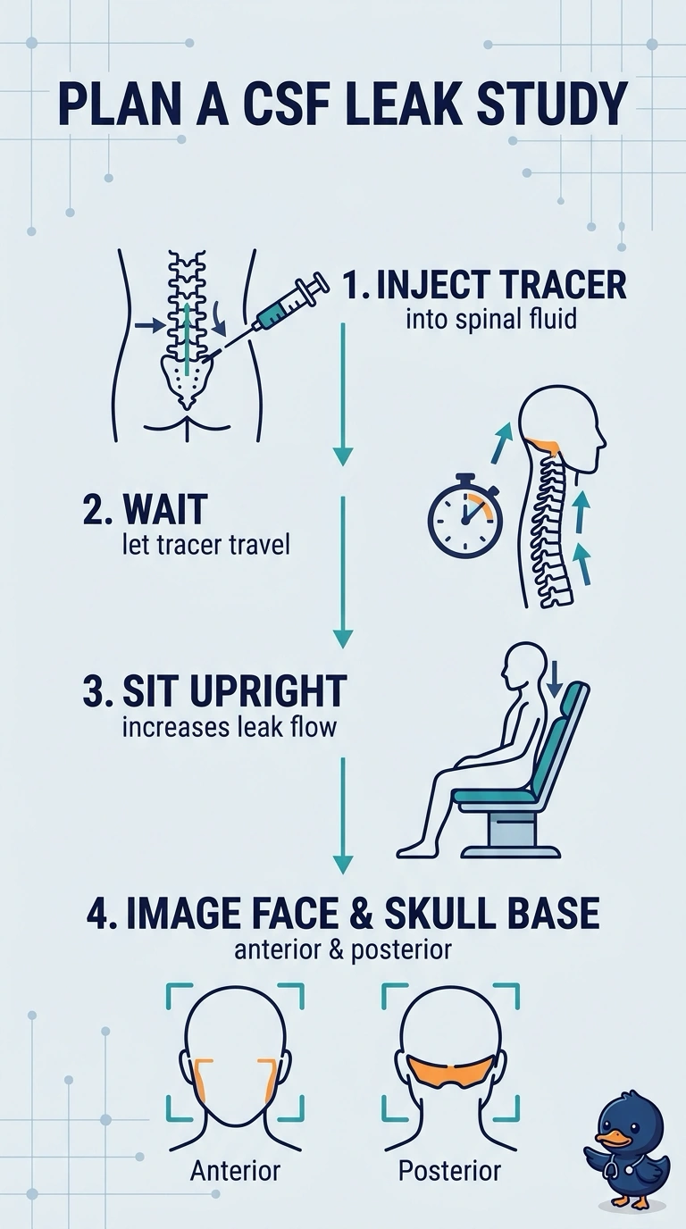
Spot on — you've caught the two things that matter, because the leak only shows once tracer has traveled and the patient sits up. Delayed upright views over the face and skull base catch fluid pooling where it escapes, much like tilting a leaky jar so the drip reaches the seam. Gravity and timing are what make the upright position work.
Full Explanation
Delayed anterior and posterior imaging of the face and skull base with the patient upright is best suited to localize a cranial CSF leak in this setting. Delayed acquisition allows intrathecal tracer time to reach and escape through the defect, while upright positioning reflects the posture that worsens the patient’s discharge. Imaging the relevant anatomy in two projections can help identify the site of egress rather than merely establish that tracer has reached the nose. Face-and-skull-base images obtained during the first hour are appropriately targeted and upright, but may be too early for tracer to accumulate at the leak; the several-hour imaging window is a reason to use delayed views. Delayed face-and-skull-base images obtained supine provide suitable timing and anatomic coverage, but do not reproduce the upright condition associated with this patient’s symptoms and may fail to demonstrate posture-dependent leakage. Delayed upright whole-head planar images can show cranial tracer distribution and nasal passage, but broad coverage is less focused on the skull base and may demonstrate nasal activity without localizing the escape site. Exam caveat: tracer reaching the nasal cavity confirms passage but does not, by itself, pinpoint the defect. Practical check: confirm the acquisition protocol includes delayed, upright views that adequately cover the face and skull base in anterior and posterior projections.