
A 71-year-old with a hip prosthesis has six months of pain and a raised inflammatory marker after revision surgery, so reactive marrow is likely around the implant. The physician orders an indium-111 labeled white cell study, and the plan must use collimation matched to the isotope, capture delayed uptake, and allow comparison with normal marrow. Which imaging plan best supports the diagnosis of chronic prosthetic infection?
Select an answer to reveal the explanation.
Short Explanation
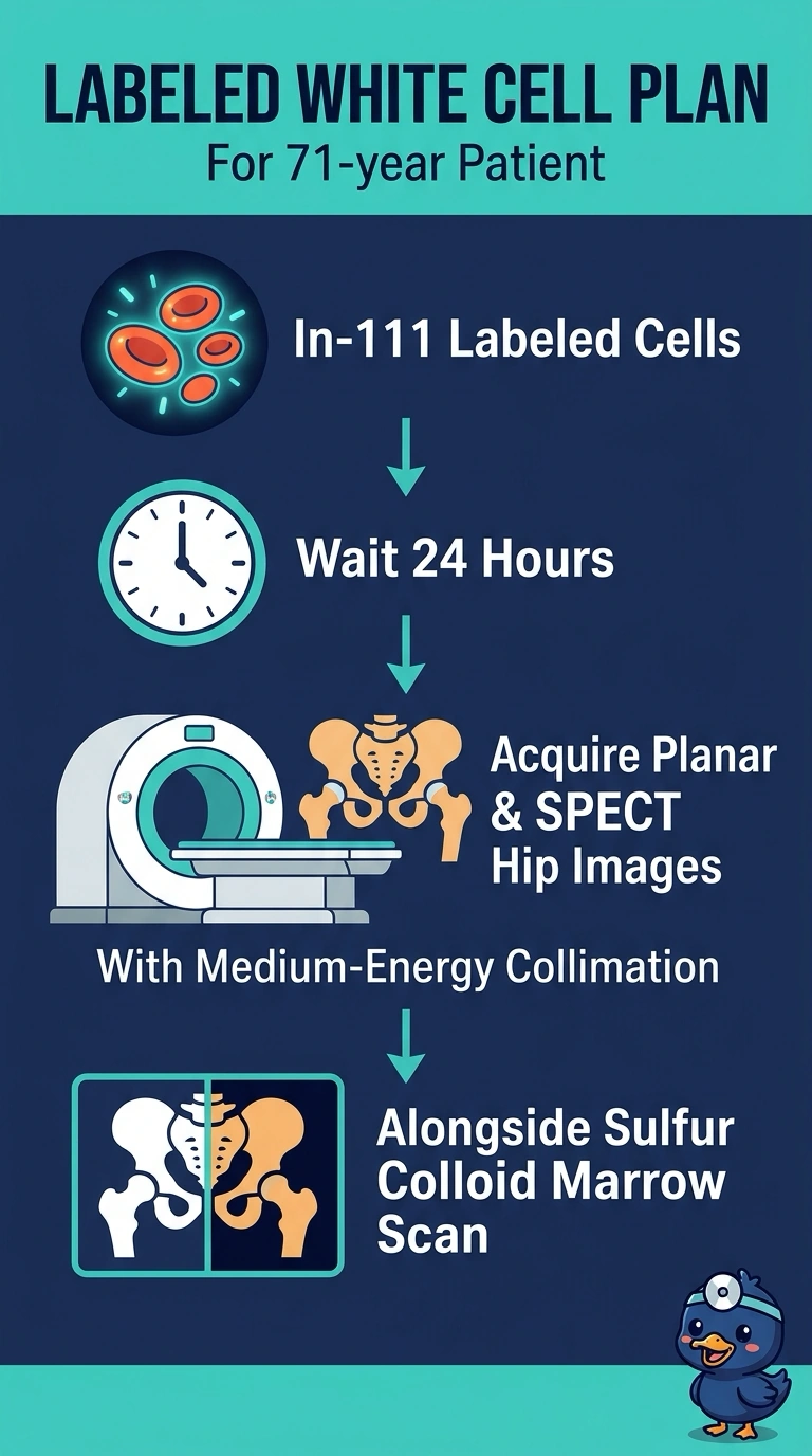
You match indium-111 with medium-energy collimation, wait until 24 hours for delayed uptake, and compare with sulfur colloid to map reactive marrow. That paired view helps you distinguish infection from marrow activity around the implant. If you change any one of those details, the uptake is harder to interpret.
Full Explanation
Indium-111 emits photons at two fairly high energies, so you need collimator septa thick enough to stop them from leaking through; medium-energy collimation preserves image quality, whereas low-energy collimation lets photons pass through and can make focal uptake look deceptively uniform. Chronic prosthetic infection accumulates white cells slowly, so imaging at 24 hours gives labeled cells time to migrate into the focus while background blood-pool activity clears. An image at 4 hours can undersell the finding because tracer has not yet concentrated. Since revision surgery can leave reactive marrow around the implant, you need to compare labeled-cell uptake with marrow distribution: sulfur colloid shows where marrow is, helping you interpret a match or mismatch. A whole-body bone scan maps osteoblastic turnover rather than marrow, so it cannot make that comparison or separate infection from a normal marrow response. Exam caveat: focus on matching the isotope, delayed uptake, and marrow comparison rather than treating the time point alone as diagnostic. Practical check: verify that medium-energy collimation, 24-hour imaging, and the sulfur colloid marrow study are documented before the patient leaves.