
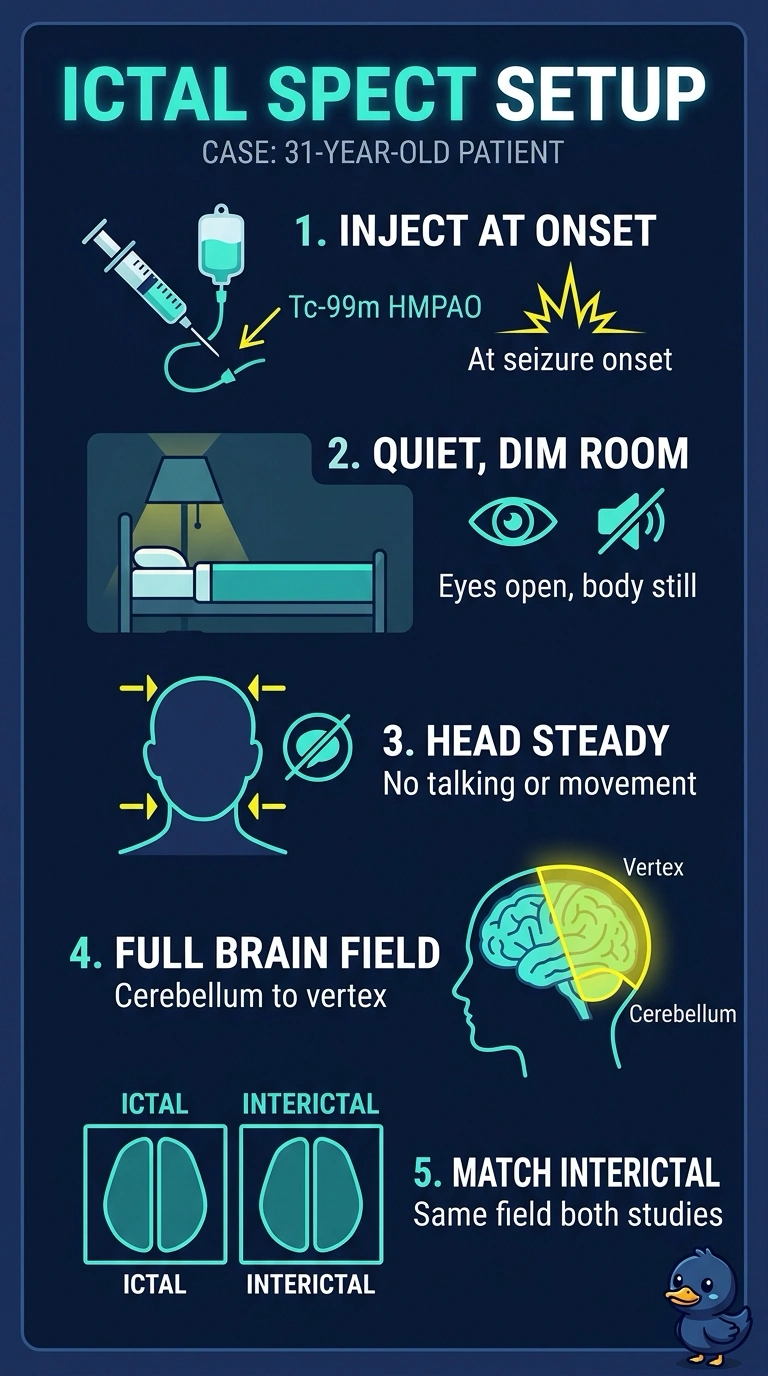
A 31-year-old with drug-resistant seizures is admitted so ictal brain perfusion SPECT can be compared with the interictal study. The tracer must go in during the seizure itself, the surroundings shape regional uptake, and the final image set must match the interictal field. Which preparation plan best preserves the ictal perfusion pattern?
Select an answer to reveal the explanation.
Short Explanation
An ictal injection captures perfusion during the seizure, which is the point of the study. Dim light, quiet, and eyes open help keep regional uptake conditions consistent for comparison with the interictal study. Miss one piece and you may capture the wrong moment or an inconsistent map.
Full Explanation
Tc-99m HMPAO crosses the blood-brain barrier and is retained in brain tissue in relation to perfusion around the time of injection, so an injection during the seizure can capture the ictal hyperperfusion pattern. Injecting after the seizure ends records the postictal period instead, when perfusion may have fallen or shifted. Regional uptake also depends on the patient's state; lighting, eye state, talking, and movement can affect the pattern, so a quiet, dim, eyes-open condition helps make the interictal comparison consistent. A restricted acquisition field fails the comparison requirement, since both studies should cover the same anatomy from cerebellum to vertex. Image acquisition can be delayed after injection because the tracer is retained in brain tissue. Verify injection timing against the seizure log and confirm both studies use comparable head positioning and reconstruction before comparison.