
A 69-year-old with heart failure and suspected cardiac amyloidosis is injected with Tc-99m PYP. At 1 hour, planar images show activity over the left heart with blood pool, sternum, and ribs overlapping, plus a bright liver. The reader must rule out blood pool before calling the study cardiac. Which acquisition best proves the uptake is truly myocardial?
Select an answer to reveal the explanation.
Short Explanation
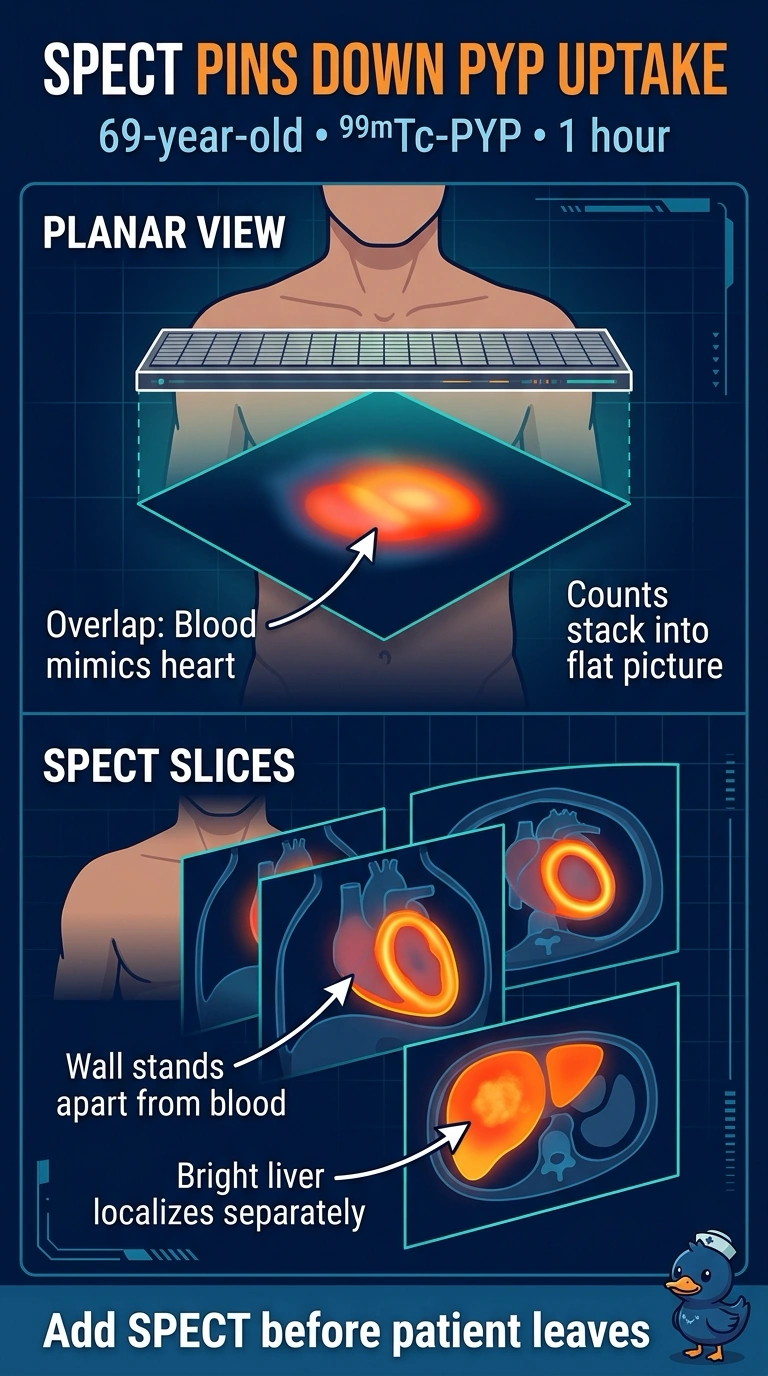
Planar imaging shows you where counts are, but not how deep they sit, and that's the problem here. SPECT slices the heart so wall uptake stands apart from chamber blood, which makes SPECT correlation your tie-breaker.
Full Explanation
Planar imaging is a projection, so it flattens the three-dimensional distribution of tracer onto a single detector plane and loses depth in the process. Counts in the ventricular blood pool, the sternum, the ribs, and the left liver lobe can all land over the left heart, and SPECT reconstructs the distribution in three axes, helping show whether tracer localizes to the myocardial wall or instead to the chamber or chest wall. A longer planar acquisition only tightens statistical noise on the same projection, so blood pool and myocardium may remain superimposed. Delayed planar imaging at three hours may reduce blood-pool activity, but it still does not provide tomographic localization. An oblique planar view changes the viewing angle rather than adding depth, so liver overlap may shrink while ventricular blood pool can still project over the myocardium. Exam caveat: heart-to-contralateral ratios are calculated from planar images, but they do not replace SPECT confirmation of myocardial uptake. Practical check: acquire SPECT before the patient leaves whenever planar appearance and the clinical story disagree.