
A 68-year-old man with treated prostate cancer has a rising PSA level. The radiologist wants the whole skeleton screened for metastatic disease and a questionable lower lumbar spine focus separated from adjacent degenerative uptake using anatomic correlation. Which imaging plan best satisfies both requirements for this tumor study?
Select an answer to reveal the explanation.
Short Explanation
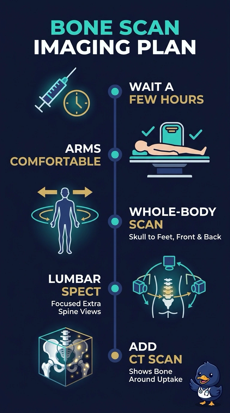
Start with whole-body planar images from skull to feet to survey the skeleton, then add lumbar SPECT/CT so you can match the suspicious uptake to its anatomy. It’s like scanning the whole map, then zooming in for street-level detail to tell degeneration from metastasis.
Full Explanation
Whole-body planar scintigraphy provides broad skeletal coverage, typically with anterior and posterior images from skull to feet. Adding SPECT across the lumbar spine provides cross-sectional localization, and the CT component supplies anatomic correlation to help assess the focus, including whether it corresponds to degenerative change or a compression fracture. A planar survey with spot views of the lumbar spine preserves skeletal coverage but does not provide the same cross-sectional localization and CT correlation. Focused SPECT/CT of the lumbar spine and pelvis with spot views of the chest, skull and ribs does not survey the entire skeleton; spot views sample only selected regions. Functional SPECT without CT can localize uptake but does not provide the requested CT-based anatomic correlation. On the exam, planar imaging surveys, SPECT localizes, and CT provides anatomic information.