
A patient with a history of breast cancer returns for a routine whole-body bone scan. The anterior and posterior images show one faint, ill-defined focus in the midsacrum that overlaps bladder activity, and the ordering physician wants better detail on that focus before therapy changes tomorrow. The patient has driven a long way, so the answer must come from today's visit. Which imaging approach should add clarity to that indeterminate sacral uptake today?
Select an answer to reveal the explanation.
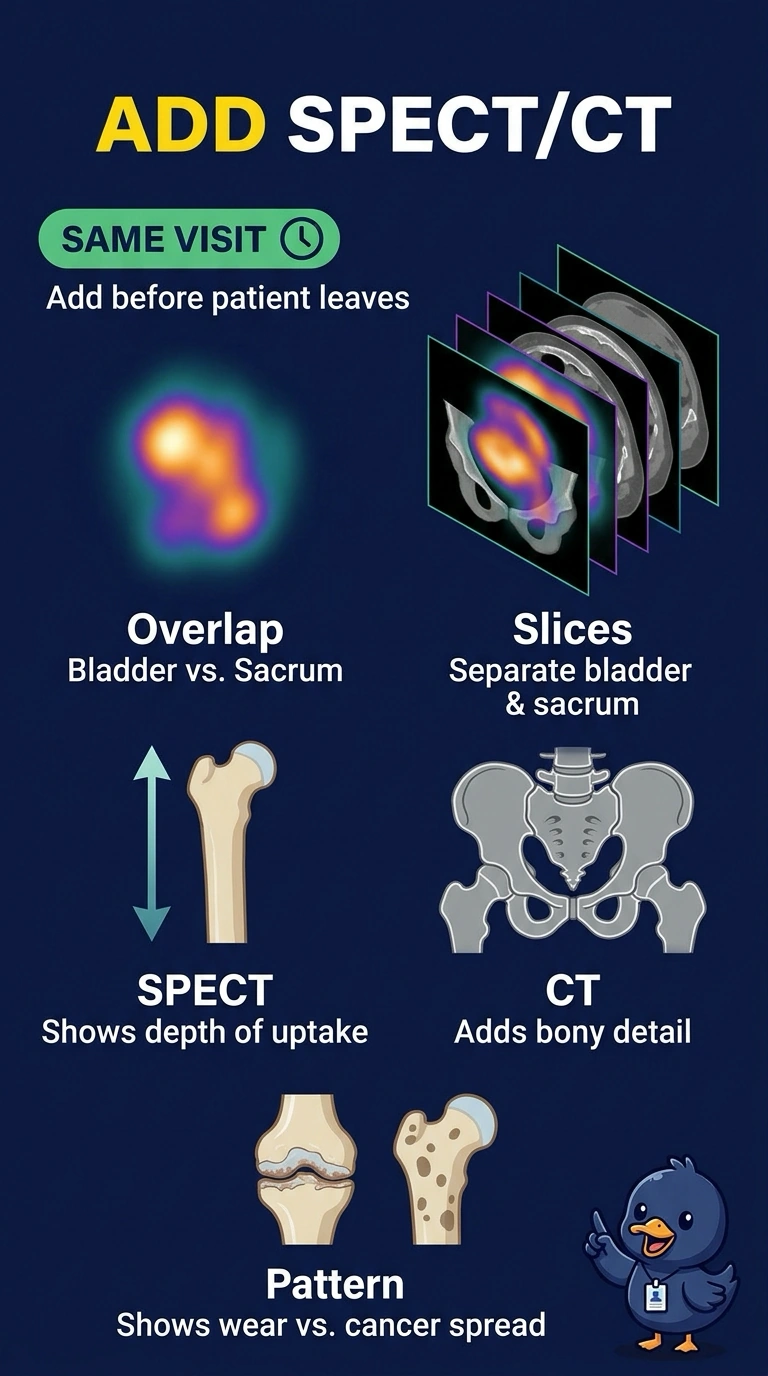
Short Explanation
You’ve spotted the problem: bladder activity overlaps the midsacrum, and longer planar imaging would leave that overlap in place. SPECT/CT separates activity by depth and adds bony anatomy, helping you assess the focus today. Think of it as moving from a stacked snapshot to cross-sectional slices.
Full Explanation
SPECT/CT is appropriate because it provides cross-sectional depth and anatomic correlation during today’s visit. Rotating detectors reconstruct slices that can separate tracer overlapping the sacrum on planar images, while the CT component shows bony anatomy; together, these details help you distinguish degenerative change from metastatic disease by location and pattern rather than brightness alone. Repeating the whole-body acquisition with a longer count time adds counts but preserves the same projection and geometric overlap, so bladder activity may still obscure the midsacral focus. Prone, zoomed static spot views provide a more focused projection, but remain two-dimensional and cannot resolve activity layered at different depths. A separate PET/CT appointment later this week could provide metabolic information, but would not meet the need for an answer from today’s visit. One exam caveat: SPECT can improve localization even without CT, and CT availability varies by suite, so recognize tomographic imaging as the essential step. As a practical check, confirm that the camera covers the sacrum and that the CT range includes the focus before the patient leaves.