
A patient with a calcified, narrowed aortic valve and worsening exertional dyspnea is scheduled for stress myocardial perfusion imaging. Cardiology documents pressure overload across the valve. Which left ventricular geometry is the expected adaptation to that load?
Select an answer to reveal the explanation.
Short Explanation
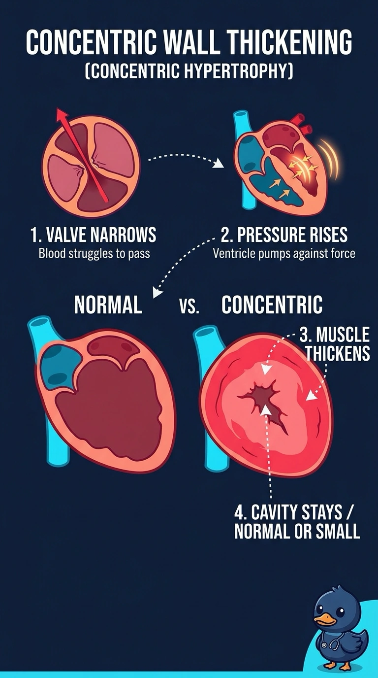
With a narrowed aortic valve, your left ventricle pushes against extra pressure, so its walls and septum thicken inward in concentric hypertrophy, usually leaving a normal-sized or smaller cavity. It’s like adding thicker walls to a pump rather than stretching its chamber, as volume overload tends to do.
Full Explanation
Pressure overload makes the ventricle generate greater systolic pressure. It commonly adapts by adding sarcomeres in parallel, producing concentric wall thickening with a cavity that is often normal in size or small. Dilatation with unchanged wall thickness describes eccentric hypertrophy, typically associated with volume overload such as regurgitation. Asymmetric septal thickening is a different pattern, commonly associated with hypertrophic cardiomyopathy. Right ventricular dilatation with septal flattening can occur with right-sided pressure overload, such as pulmonary hypertension. Long-standing pressure overload can eventually lead to ventricular dilatation and failure, so the geometry depends on the stage of disease. Gated myocardial perfusion images assess LV function and wall motion; echocardiography or cardiac MRI is generally used to characterize wall thickness and geometry.