
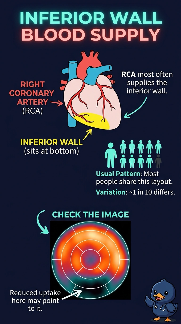
On a myocardial perfusion study the reduced tracer uptake sits in the inferior wall of the left ventricle on both stress and rest images, while the other walls fill normally. Which coronary artery most commonly supplies the territory where the uptake is reduced?
Select an answer to reveal the explanation.
Short Explanation
The inferior wall is supplied by the right coronary artery in most people, so that is where your eye goes when uptake dips at the bottom of the left ventricle. About one in ten circulation patterns shifts to left dominance, which is the wrinkle worth remembering. The three main arteries work like three delivery routes, each stocking its own neighborhood.
Full Explanation
The inferior wall of the left ventricle is the classic territory of the right coronary artery in the majority of people, which is why reduced uptake there directs attention to that vessel. Coronary dominance varies: when the circumflex gives rise to the posterior descending artery, the inferior wall is supplied by the left circumflex instead, so this is a most-common pattern rather than an absolute rule. The left anterior descending artery supplies the anterior wall and apex, so an anterior or apical defect would point to that vessel rather than to the right coronary artery. The left circumflex supplies the lateral and posterolateral segments, and in the usual pattern that territory is not the inferior wall. The left main trunk is a short parent vessel that divides into the anterior descending and circumflex arteries, so naming it does not localize a defect to any single wall. Caveat: dominance differs between patients, and an inferior defect can also reflect attenuation from the diaphragm rather than disease. Practical check: confirm the wall location on the short-axis images before naming an artery, then compare stress with rest to judge whether the finding is reversible.