
During hepatic arterial perfusion scintigraphy for a lung shunt fraction before Y-90 microsphere therapy, the technologist's Tc-99m MAA syringe has been sitting on the counter during a delay while the team secures the catheter in the planned treatment position. Planar images must support a lung-to-liver count comparison. Which preparation and acquisition sequence will produce a valid shunt fraction?
Select an answer to reveal the explanation.
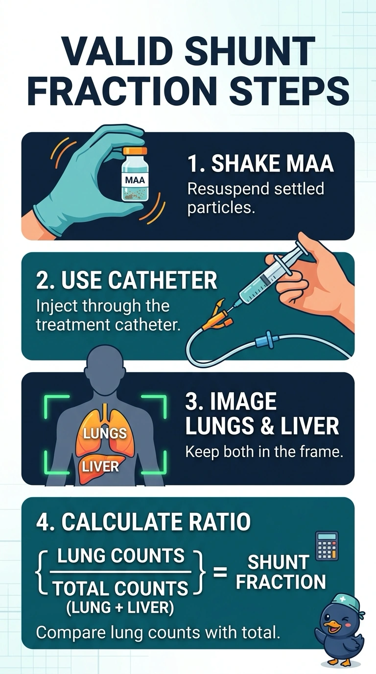
Short Explanation
You’ve covered all three bases: a resuspended dose, the treatment catheter, and images of both lungs and the liver. Settled MAA is like a snow globe that has stopped swirling, so its particles may no longer represent true flow. Resuspending the MAA right before injection is the step people often miss.
Full Explanation
Macroaggregated albumin (MAA) particles settle in the syringe within minutes, so injecting settled MAA through the treatment catheter can deliver a biased particle distribution that no longer reflects blood flow through the tumor bed and can skew the lung shunt estimate. Resuspend the dose, inject through the catheter secured in the planned treatment position, and image both lungs and the liver: the shunt fraction predicts risk for the territory therapy will treat, and its count ratio compares lung activity with total activity. Injecting settled MAA through the treatment catheter ignores sedimentation. Resuspending MAA but injecting it through a peripheral vein sends particles through a different route and does not sample hepatic trapping from the planned treatment position. Resuspending MAA, injecting through the treatment catheter, and imaging only the abdomen leaves out the lung counts needed for the ratio. One exam caveat: some protocols add anterior and posterior views or SPECT, but the comparison still requires both lungs and the liver. Practical check: swirl the syringe immediately before injection and confirm on the display that both lungs and the liver are inside the field of view.