
Myocardial tracer uptake rises with regional coronary blood flow, so perfusion imaging compares stress uptake with rest uptake. A defect with reduced stress uptake and normal rest uptake in the anterior septum and apex, with the inferior and lateral walls normal, is reversible. Which pathophysiology statement best explains that pattern?
Select an answer to reveal the explanation.
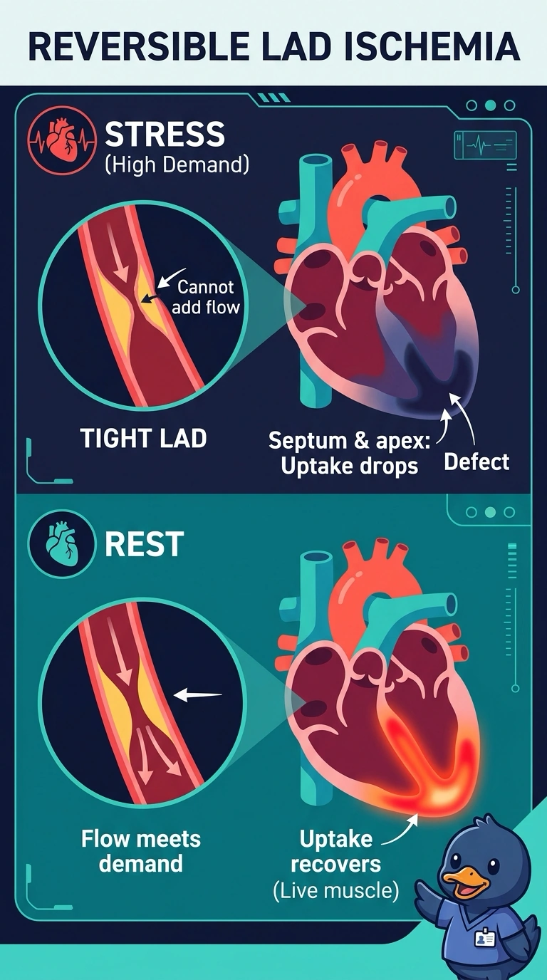
Short Explanation
Reversible means the muscle is alive and only short of blood during stress. Here the left anterior descending artery can't open up enough flow for the anterior septum and apex, so uptake falls until rest restores supply. You're seeing the classic fill-in pattern of ischemia, exactly what stress imaging is built to expose.
Full Explanation
Regional tracer uptake during stress scales with coronary blood flow, and a vessel with limited flow reserve cannot raise perfusion enough for the anterior septum and apex, so those segments take up less tracer under stress while the same viable segments fill in once rest restores flow; that reversibility is the signature of ischemia in the left anterior descending territory. Scar from a healed infarct fails the pattern because scar tissue produces a fixed defect that stays reduced at both rest and stress, contradicting the fill-in finding. Attenuation from body habitus often produces a similar reduction on rest and stress images, rather than a defect limited to stress. Right coronary demand mismatch fails because that vessel mainly serves the inferior wall, which the stated pattern shows as normal. Caveat: diaphragmatic attenuation is the classic false inferior defect, so compare rest and stress images before calling a segment abnormal. Practical check: review gated wall motion; reversibly ischemic segments often contract normally at rest.