
A patient with known breast cancer reports new focal low-back pain after finishing therapy. Whole-body planar bone imaging shows no abnormal uptake, and lumbar radiographs show only mild degenerative change. Which added acquisition best localizes the symptomatic lesion?
Select an answer to reveal the explanation.
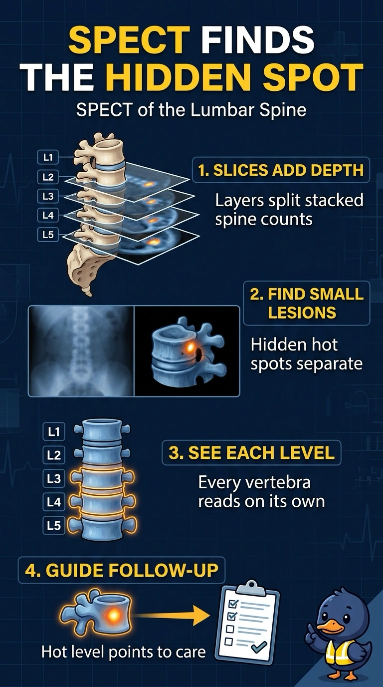
Short Explanation
When flat pictures look clean but your pain is real, SPECT helps separate overlapping activity in the spine. Cross-sectional views show which vertebral level is active. Tomography adds the depth detail planar images cannot provide.
Full Explanation
SPECT produces cross-sectional slices, separating activity that overlaps in summed anterior or posterior planar views by depth and vertebral level. This added dimension helps localize a lesion that a whole-body survey may not reveal, especially in the posterior elements, where normal activity can obscure a focus. A limited delayed-only lumbar study fails conceptually because it repeats planar imaging, the same geometry that was already negative, without adding depth information. Three-phase lumbar imaging fails because flow and blood-pool phases assess perfusion, which is useful for questions such as infection or complex regional pain syndrome, whereas this patient needs detection and localization of a possible malignancy-related lesion. Repeating whole-body planar imaging fails because that survey already showed no abnormal uptake; another sweep with the same technique is unlikely to explain focal pain. Exam caveat: radiographs showing only mild degenerative change do not exclude a metabolically active lesion. Practical check: when symptoms and planar findings disagree, consider tomography of the painful region before accepting a negative survey.