
While reviewing a myocardial perfusion SPECT study, you note that the camera logged a shift in the patient's position partway through acquisition, and the summed images show displaced subdiaphragmatic activity over the inferior wall. Which data processing approach should be applied to correct this dataset?
Select an answer to reveal the explanation.
Short Explanation
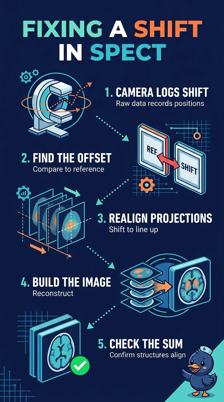
Use motion correction to realign the projection data to one reference position before reconstruction, like lining up shifted frames in a video. Sharper filtering, CT-based attenuation correction, and ECG gating address other issues—not your patient’s position shift.
Full Explanation
Motion correction is appropriate because the camera recorded a change in the patient’s position during acquisition. Realigning the affected projection data to a common reference position before reconstruction corrects geometric misregistration between the inferior wall and the displaced subdiaphragmatic activity. Applying a sharper reconstruction filter can change image resolution and noise, but it cannot realign projection data or correct movement. CT-based attenuation correction compensates for photon absorption in tissue rather than patient displacement; if the attenuation map is mismatched to the emission data, it can introduce artifacts. ECG gating sorts counts by cardiac phase to assess function across the cardiac cycle, but it does not correct whole-body translation. An exam caveat is that motion correction depends on the shift being identifiable and correctable; large or poorly characterized motion may leave residual misregistration. As a practical check, review the summed cine images and motion information before reconstruction to confirm that the projections can be aligned to a consistent reference position.