
A patient with biochemically confirmed primary hyperparathyroidism has a nondiagnostic neck ultrasound, and the surgeon plans a minimally invasive, image-guided exploration and wants the gland's precise side, level and depth. Which parathyroid imaging plan best satisfies that localization request?
Select an answer to reveal the explanation.
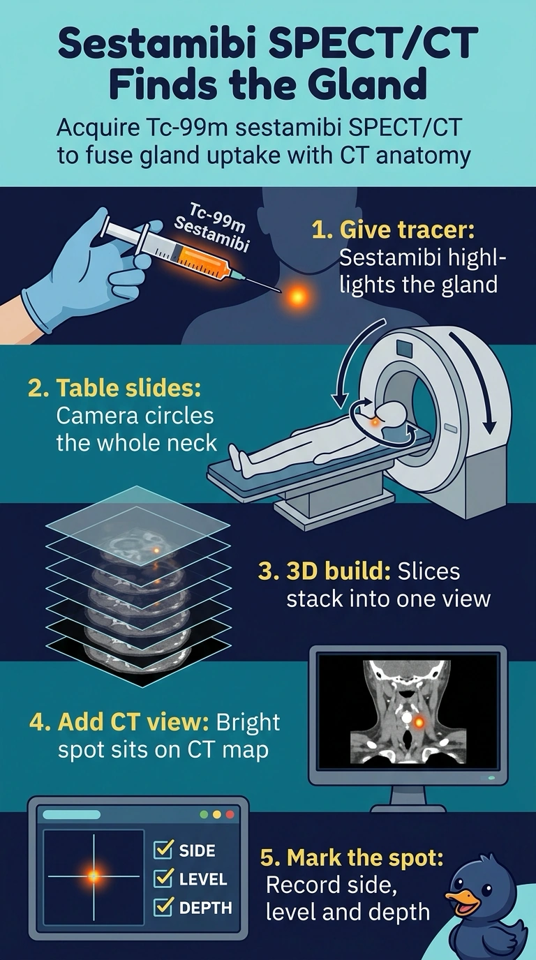
Short Explanation
The surgeon is really asking for coordinates, not just a bright spot. Sestamibi marks the overactive tissue, and the CT half of SPECT/CT puts it on a map you can plan an incision around. It's fusion that turns a hot spot into a surgical address.
Full Explanation
Sestamibi is retained by mitochondria-rich parathyroid tissue, and adding SPECT/CT fuses that uptake with cross-sectional anatomy so the gland is reported by side, level and depth, exactly the information an image-guided minimally invasive exploration needs. Dual-phase planar imaging relies on washout differences to separate gland from thyroid, but the planar approach projects everything onto one plane, cannot supply depth and cannot reliably distinguish a deep adenoma from overlying thyroid. Subtraction imaging improves functional contrast between thyroid and parathyroid, yet the subtraction technique still yields a two-dimensional answer without the axial coordinates the surgeon asked for. Pinhole spot views magnify small neck structures, but the magnification approach has no tomographic reference and can miss a gland behind the sternum. Caveat: when a question only asks how to detect an adenoma, dual-phase or subtraction answers may both be defensible, so localization wording is what points to fusion imaging. Practical check: confirm the report names the gland relative to the trachea and a vertebral or clavicle level.