
A busy nuclear medicine department schedules two studies back to back: a young patient with recurrent painless rectal bleeding and suspected ectopic gastric mucosa whose focus may appear only after several minutes of circulation, and an older inpatient with recurrent melena after inconclusive endoscopy whose bleeding is slow and intermittent. Which pair of gastrointestinal studies fits both referrals?
Select an answer to reveal the explanation.
Short Explanation
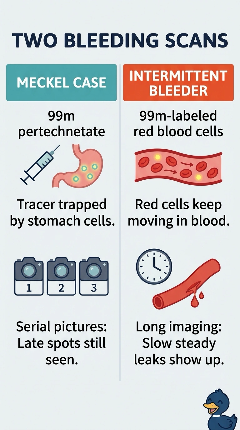
For suspected ectopic gastric mucosa, you use Tc-99m pertechnetate with serial images, since functioning stomach cells take it up and the focus may take time to appear. For slow, intermittent bleeding, Tc-99m-labeled red blood cells act like a circulating tracker, helping you catch bleeding that comes and goes.
Full Explanation
Technetium-99m pertechnetate is taken up by functioning gastric mucosal cells through the same anionic transport used by thyroid and salivary tissue, so a Meckel diverticulum containing ectopic gastric mucosa appears as a focal area of activity that may declare itself only after the tracer has circulated, which is why serial abdominal images are part of the plan. Technetium-99m-labeled red blood cells suit the second referral because the tagged cells remain in the vascular space and recirculate, letting a slow intermittent bleed be sampled repeatedly over a prolonged study with greater sensitivity for slow bleeding than a single quick pass. The reticuloendothelial colloid approach fails for the two referrals in different ways: sulfur colloid is cleared rapidly by phagocytes and is best suited to active bleeding, and it is not taken up by gastric mucosa, so it cannot identify a Meckel diverticulum. The immediate-images-only planar approach with pertechnetate fails because early truncation misses ectopic uptake that was not yet visible. An exam caveat is that urinary activity in the bladder and kidneys can mask a focus low in the abdomen. A practical check is confirming the eluate is pure pertechnetate and asking the patient to void just before imaging starts.