
A patient with a joint replacement has had fever and rising inflammatory markers for several weeks, and earlier surgery changed the marrow pattern around the prosthesis. The physician must tell true infection apart from normal marrow uptake there. Which imaging plan gives the most specific result?
Select an answer to reveal the explanation.
Short Explanation
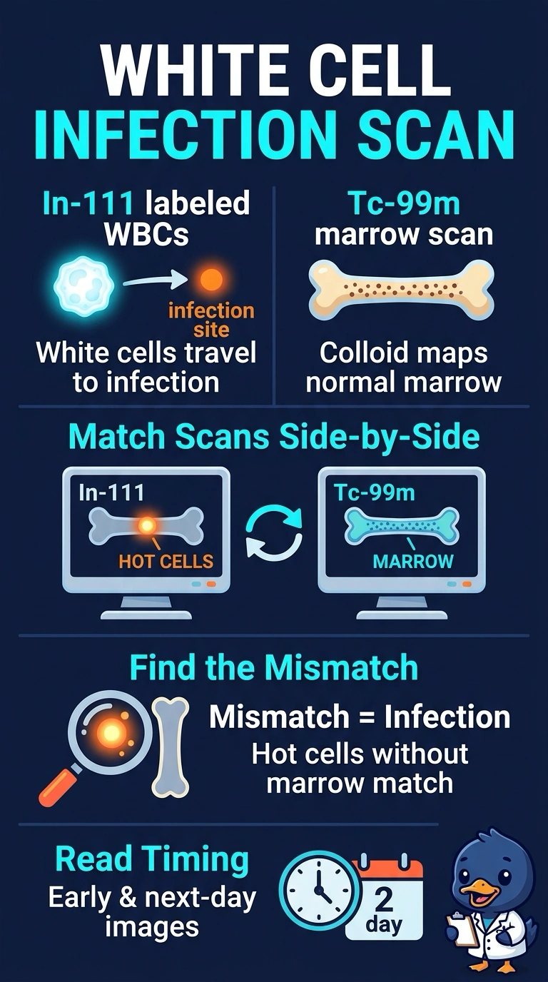
Around a prosthesis, normal marrow can light up as brightly as an infected focus, so one study alone can mislead you. You're pairing the labeled white cells with a marrow scan so both distributions sit side by side. It's the mismatch that tells you where infection actually is.
Full Explanation
Labeled white cells accumulate where chemotaxis draws them, but reticuloendothelial marrow also holds cells, and surgery that redistributes marrow makes that physiologic uptake unpredictable around a prosthesis. Adding a sulfur colloid study maps where normal marrow sits, so the read becomes a comparison: white cell uptake without a matching marrow focus points to infection, while matched uptake points to marrow. A white cell plan alone lacks that reference frame and invites false positives wherever marrow was disturbed. A bone scan alone reads osteoblastic turnover, which rises with fracture, loosening and postoperative healing just as it does with infection, so it cannot separate infection from repair. An alternate labeled cell agent improves resolution and scheduling convenience, but changing agents does nothing about the marrow comparison the case demands. Exam caveat: the order and timing of the two studies vary by department, and some add SPECT/CT for registration, so carry the comparison concept instead of a schedule. Practical check: verify labeling quality, then review both studies side by side before calling any focus infected.