
A patient with primary hyperparathyroidism is referred for localization before a minimally invasive parathyroidectomy. Neck ultrasound shows a thyroid nodule in the same area, with no recent iodine exposure reported. The department offers dual-phase planar sestamibi, same-day sestamibi with pertechnetate subtraction, sestamibi SPECT without CT, and side-by-side comparison of sestamibi and pertechnetate thyroid images. Which acquisition plan best separates thyroid uptake from hyperfunctioning parathyroid tissue?
Select an answer to reveal the explanation.
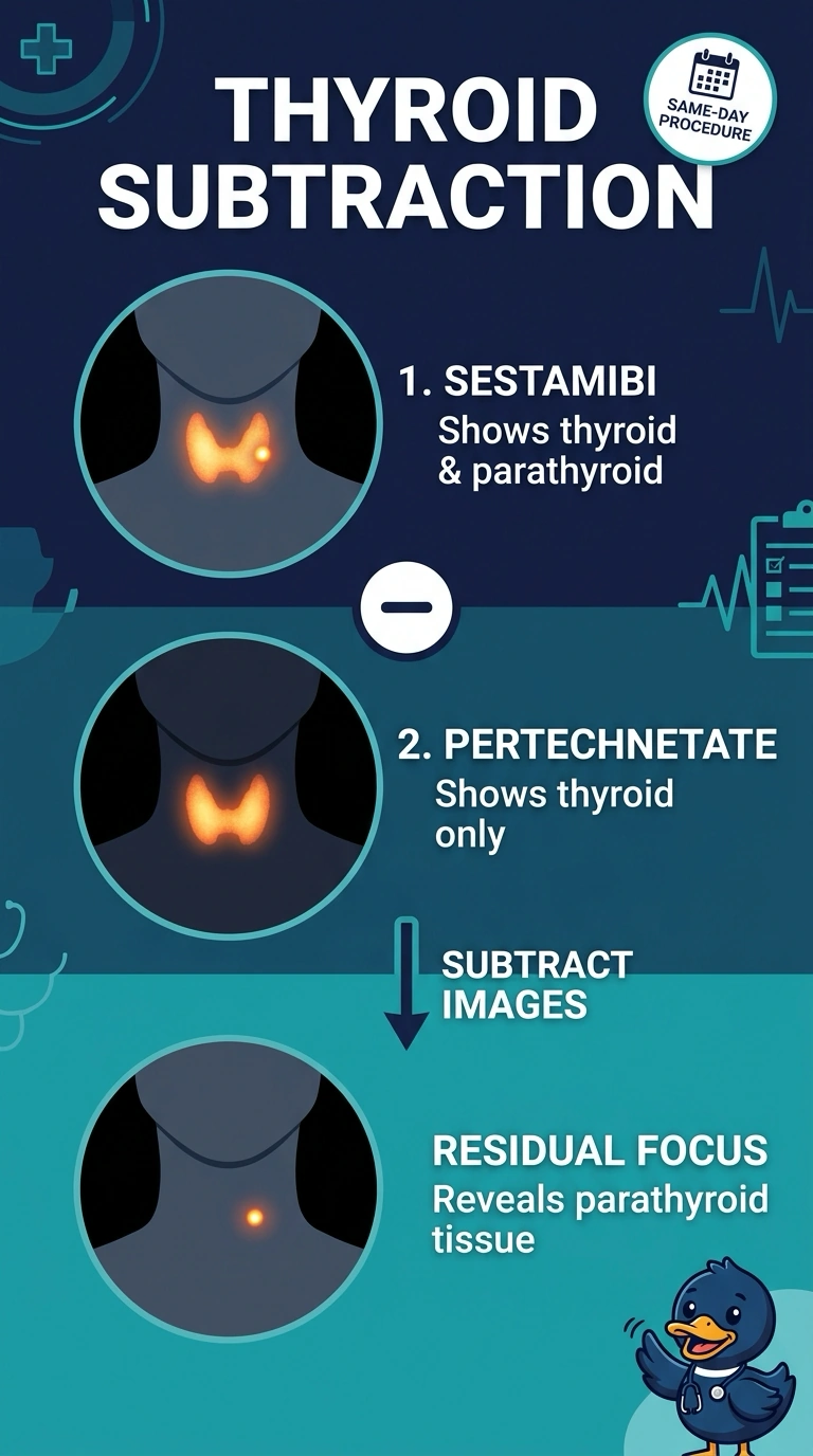
Short Explanation
You picked the approach that estimates and removes the thyroid contribution rather than relying on delayed washout. Pertechnetate subtraction can help reveal a parathyroid focus, though it does not guarantee that every thyroid nodule will be completely removed from the image.
Full Explanation
Sestamibi is taken up by both thyroid tissue and hyperfunctioning parathyroid tissue, so a nodule that overlaps the gland can complicate interpretation. Pertechnetate depicts thyroid uptake; subtracting a suitably normalized pertechnetate image from the sestamibi image can reduce the thyroid contribution and help reveal a parathyroid focus. The dual-phase washout approach depends on differential clearance, which can be unreliable, particularly when a thyroid nodule retains sestamibi. Sestamibi SPECT without CT adds depth information but does not distinguish thyroid from parathyroid tissue. Side-by-side visual comparison leaves both count patterns present and may be difficult to interpret when the tissues overlap. Subtraction results depend on acquisition and processing, and residual activity or subtraction artifacts may remain. Confirm the department's subtraction protocol and use consistent positioning and acquisition parameters.