
A patient with raised parathyroid hormone and hypercalcemia has a negative neck ultrasound but is booked for a minimally invasive parathyroidectomy. The surgeon needs the overactive tissue located in three dimensions, covered down into the chest, and distinguished from the thyroid. Which parathyroid localization plan meets those requirements?
Select an answer to reveal the explanation.
Short Explanation
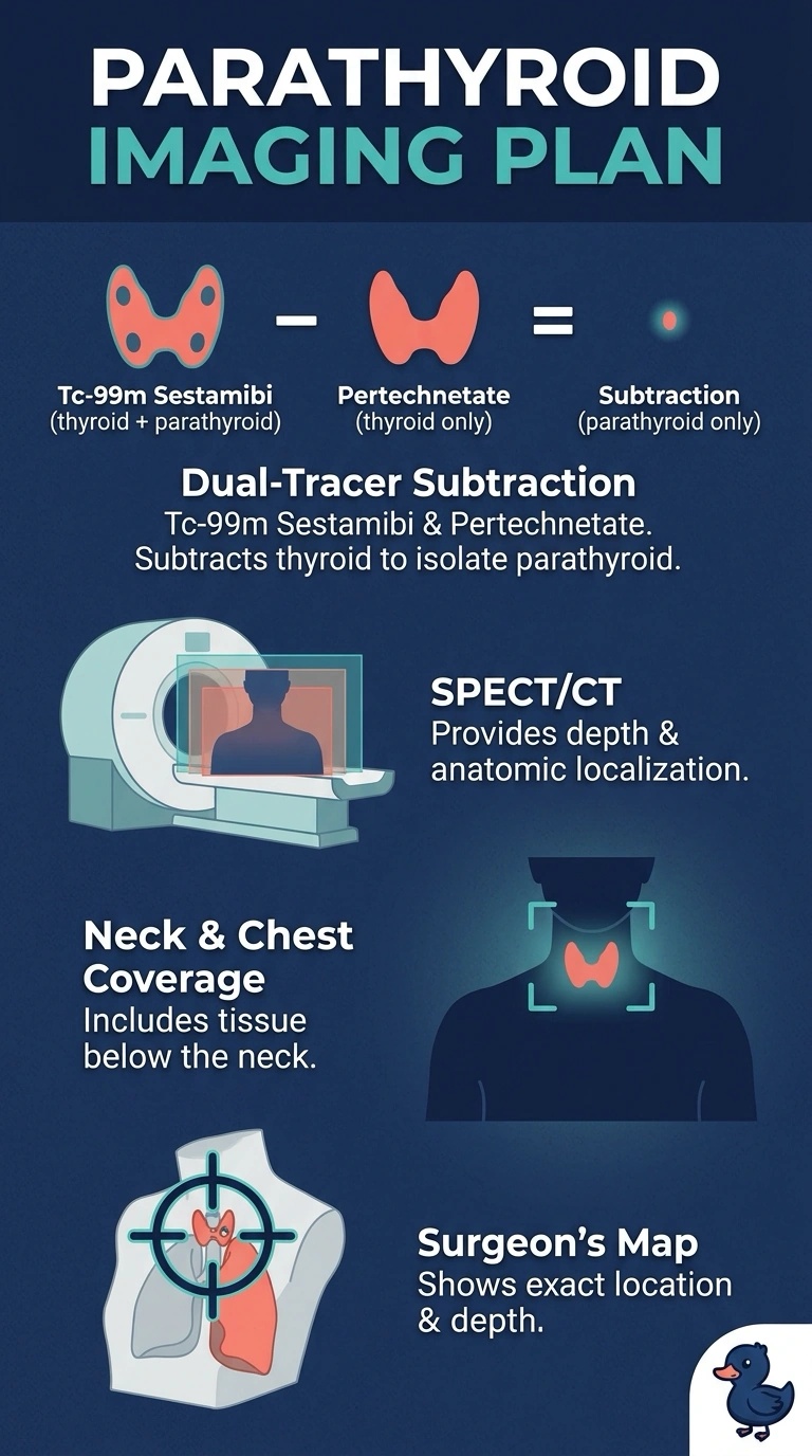
You can distinguish the overactive parathyroid tissue from the thyroid with dual-tracer subtraction using sestamibi and thyroid-seeking pertechnetate. Add SPECT/CT of your neck and chest for a three-dimensional surgical map that can include ectopic tissue below the neck.
Full Explanation
Dual-tracer Tc-99m sestamibi/pertechnetate subtraction with SPECT/CT covering the neck and chest meets all three needs: subtraction helps distinguish hyperfunctioning parathyroid tissue from thyroid activity, SPECT/CT provides three-dimensional localization, and chest coverage can reveal ectopic tissue below the neck. Dual-phase planar sestamibi views of the neck and chest can use tracer washout, but planar images do not provide tomographic three-dimensional localization or reliable depth information. Early sestamibi SPECT/CT of the neck and chest provides three-dimensional anatomy and broad coverage, but without thyroid subtraction, thyroid activity can make the adenoma harder to distinguish. Early and delayed sestamibi SPECT/CT with thyroid subtraction offers thyroid separation and three-dimensional localization, but neck-only coverage can miss an overactive gland in the chest. An exam caveat: a negative ultrasound does not exclude primary hyperparathyroidism or an ectopic adenoma; localization imaging guides surgery rather than establishing the biochemical diagnosis. In practice, confirm that the protocol explicitly includes subtraction, SPECT/CT, and chest coverage, and review the field of view and image quality with nuclear medicine before surgery.