
An endocrine surgeon plans a minimally focused neck incision for a patient with biochemically confirmed primary hyperparathyroidism and an unremarkable ultrasound, with possible mediastinal exploration. The study must show parathyroid function and provide anatomic guidance for the incision. Which parathyroid imaging plan best meets the surgical request?
Select an answer to reveal the explanation.
Short Explanation
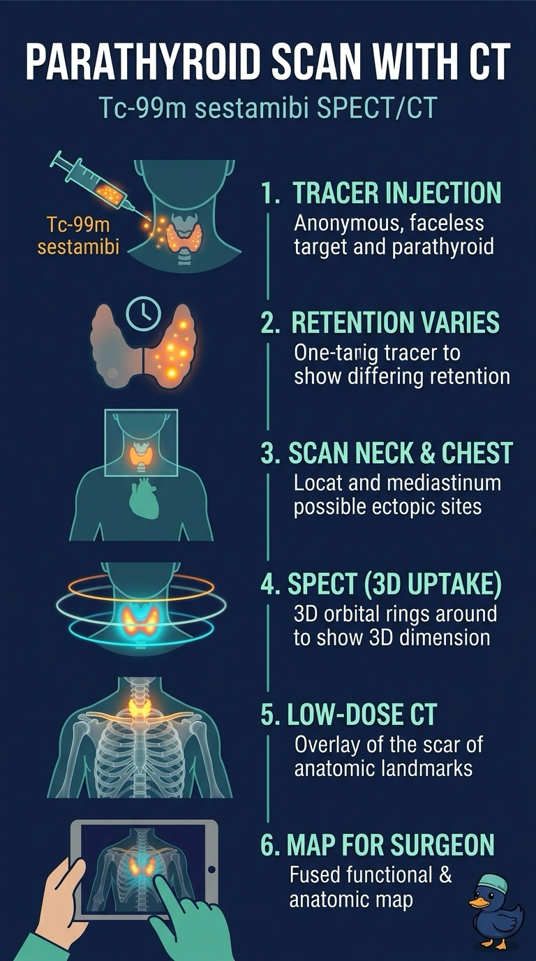
With your normal ultrasound and possible mediastinal exploration, Tc-99m sestamibi SPECT/CT scans your neck and mediastinum to show active parathyroid tissue alongside CT anatomy. It’s like pinning a glowing trail onto a map, giving your surgeon functional information and landmarks to guide the incision.
Full Explanation
Tc-99m sestamibi SPECT/CT covering the neck and mediastinum best meets the request: sestamibi uptake provides functional localization, while fused CT supplies anatomic landmarks to guide a focused incision and can localize ectopic mediastinal glands. Neck-only sestamibi SPECT/CT provides fused functional and anatomic information, but its limited coverage could miss a mediastinal target when exploration may be needed. Sestamibi SPECT of the neck and mediastinum covers both relevant regions and maps tracer distribution, but without fused CT it lacks the anatomic detail requested for incision planning. Four-dimensional CT of the neck and mediastinum offers detailed anatomic localization, but depicts enhancement and washout rather than directly demonstrating parathyroid tracer function, so it does not meet the functional-imaging requirement. A negative or nonlocalizing study does not negate biochemically established primary hyperparathyroidism; imaging is for localization and operative planning, not diagnosis. As a practical check, confirm that the acquisition protocol explicitly includes the mediastinum and that the fused SPECT/CT images are reviewed for concordance between focal uptake and the anatomic target.