
On a 64-year-old's PET/CT for lymphoma restaging, a bright crescent of uptake appears along the diaphragm on the attenuation-corrected image but is absent from the non-corrected emission image, and the CT liver dome sits lower than the PET liver dome. Which CT acquisition technique should you verify before the next chest study to reduce the chance of this mismatch recurring?
Select an answer to reveal the explanation.
Short Explanation
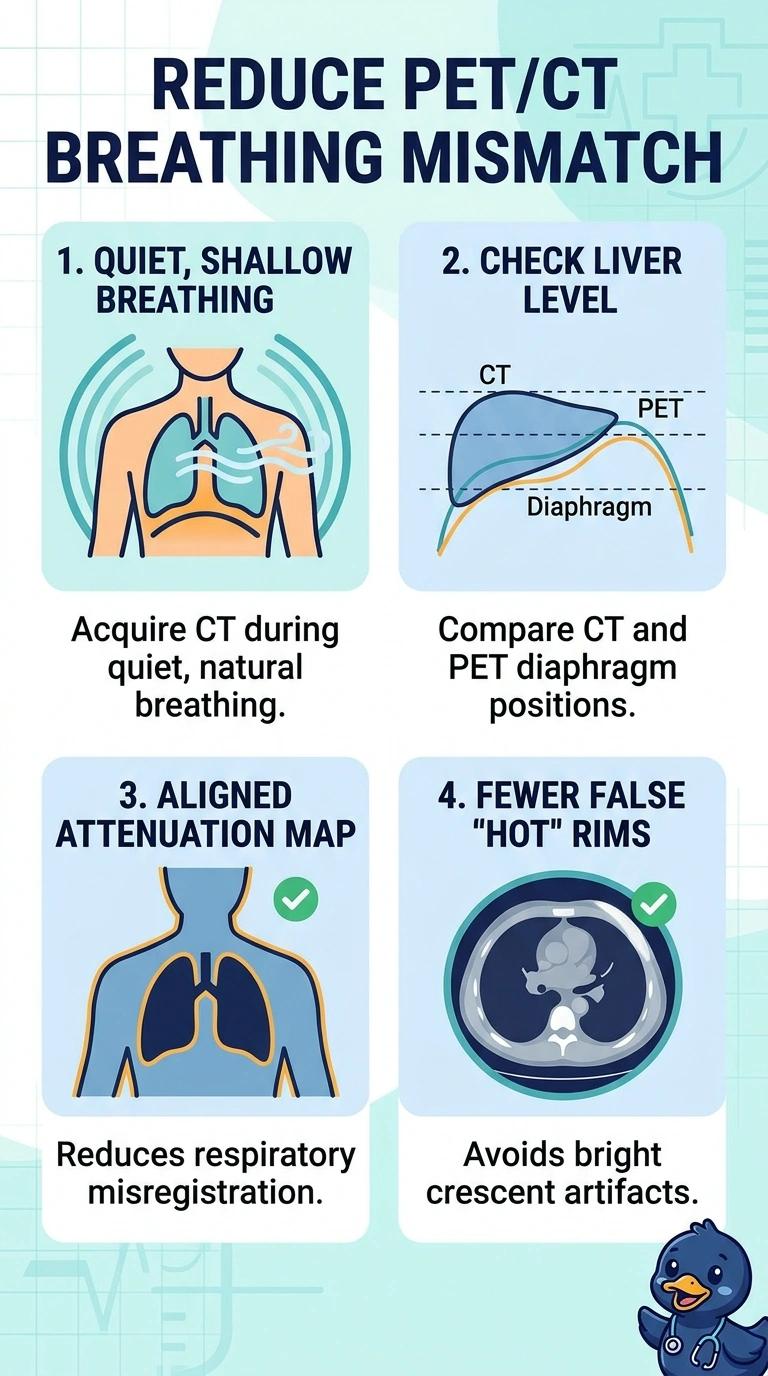
Your CT and PET should capture the diaphragm in the same breathing position—like lining up two snapshots—so check that the attenuation-correction CT uses quiet, shallow breathing. This can reduce respiratory misregistration and the false rim of uptake on corrected images.
Full Explanation
The bright crescent seen only after attenuation correction, together with the lower liver dome on CT than on PET, points to respiratory misregistration: the CT attenuation map places the diaphragm and liver differently from the time-averaged PET emission data. A low-dose CT acquired during quiet, shallow breathing is therefore the appropriate technique to verify, because it more closely matches the free-breathing PET acquisition and reduces the chance that the CT map will misclassify tissue near the moving diaphragm. A deep-inspiration breath-hold CT is a common way to obtain a sharp chest image, but its expanded lungs and elevated diaphragm do not represent the free-breathing PET position. An end-expiratory breath-hold can also reduce motion and give consistent anatomy, yet a held respiratory phase still differs from PET averaged across ongoing breaths. Respiratory-gated CT can localize anatomy at a selected phase, but gating CT alone does not make that phase correspond to an ungated, free-breathing PET acquisition. The exam caveat is that attenuation-correction artifacts may create or exaggerate apparent uptake and should not automatically be interpreted as disease. In practice, check the CT breathing instruction and review the fused images alongside the non-attenuation-corrected PET before acting on a focal finding.