
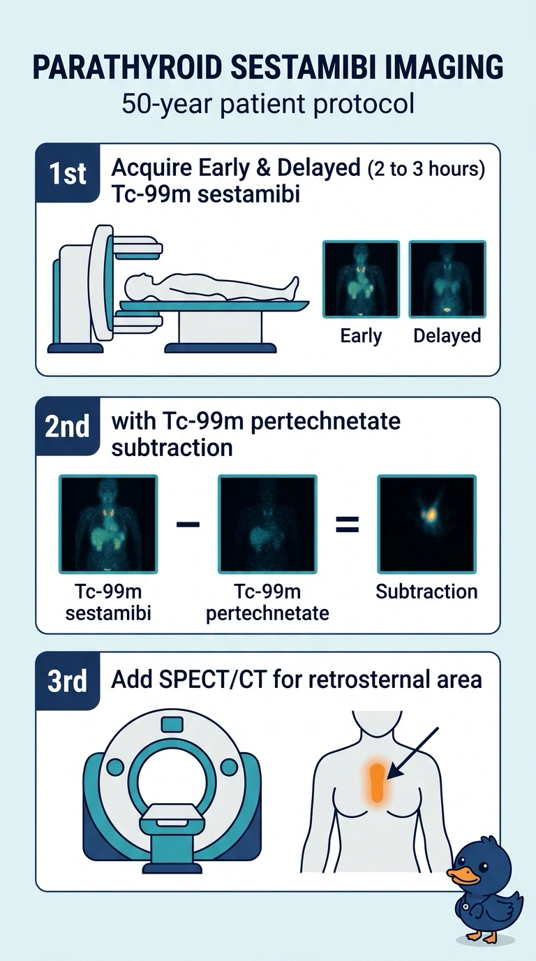
A 50-year-old outpatient with biochemically confirmed hyperparathyroidism needs preoperative localization. Neck ultrasound shows a thyroid nodule that could retain tracer, the surgeon wants early and delayed sets to judge washout, and the gland may sit behind the sternum. Which imaging plan best localizes the abnormal parathyroid?
Select an answer to reveal the explanation.
Short Explanation
You’ll compare early and delayed sestamibi images like checking which tissue lets go of the tracer more slowly, while pertechnetate imaging helps subtract thyroid activity from a nodule. Add SPECT/CT to pinpoint the gland’s location, especially if it’s behind the sternum.
Full Explanation
Two-phase sestamibi imaging can show differences in tracer washout between thyroid and hyperfunctioning parathyroid tissue, although washout patterns are variable and are not definitive by themselves. A pertechnetate thyroid image can be used for subtraction to help distinguish thyroid uptake from sestamibi activity; it is not a thyroid-only anatomic map, and a nodule’s uptake patterns may complicate interpretation. SPECT/CT adds anatomic localization and can help localize a retrosternal focus. Planar images may include the mediastinum, but provide less depth and anatomic localization than SPECT/CT. A single early acquisition does not provide the requested washout comparison. Dual-phase imaging without subtraction may still be informative, but is less suited to this scenario because thyroid nodular uptake could confound interpretation. Sensitivity can be lower with multigland disease and small glands, so a negative study does not rule out hyperparathyroidism.