
A 58-year-old outpatient with diabetes and early satiety is booked for a solid-meal gastric emptying study to check for gastroparesis. He has fasted since midnight and the order calls for a four-hour protocol. Which meal timing and imaging schedule suits this gastric emptying study?
Select an answer to reveal the explanation.
Short Explanation
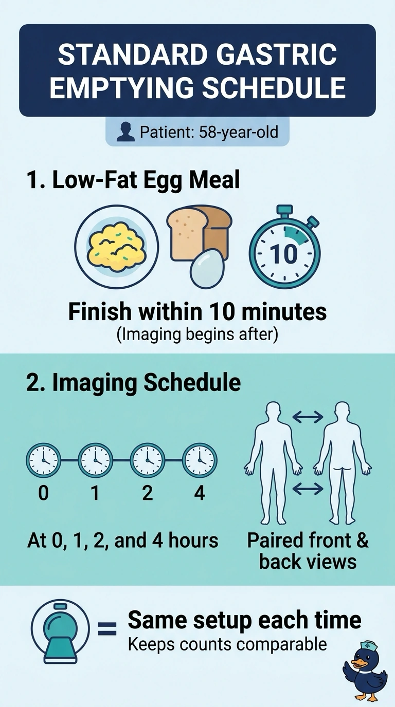
You should finish the standard low-fat egg meal within 10 minutes, then get paired anterior and posterior images at 0, 1, 2, and 4 hours. These paired views keep the measurements comparable at every time point—like checking a race at each mile, not just the first two.
Full Explanation
The protocol is built on reproducibility: a low-fat egg meal finished within ten minutes delivers a fixed solid burden, and Tc-99m sulfur colloid remains trapped in the cooked protein, so counts over the stomach represent food that has not emptied yet. Paired anterior and posterior views let you take a geometric mean, which reduces the effect of changing stomach depth, and the 0, 1, 2, and 4 hour points capture late retention that can indicate delayed emptying. A single-detector count-rate setup does not provide the standard paired-view geometric-mean measurement. An abbreviated schedule ending at two hours does not collect the four-hour measurement. A liquid test meal assesses liquid-phase emptying, which is physiologically different and is not a substitute for this solid-phase study. Some departments may use a shorter protocol when the clinician accepts limited information, so follow the written procedure. Record the meal's start and finish times before the patient leaves.