
A 55-year-old with long-standing diabetes is scheduled for solid-meal gastric emptying scintigraphy. The patient arrives after an overnight fast, finishes the labeled egg meal within the allowed time, and feels much better by the 2-hour image, so the nurse suggests ending the study there. Which imaging schedule best answers the referring clinician's question about delayed emptying?
Select an answer to reveal the explanation.
Short Explanation
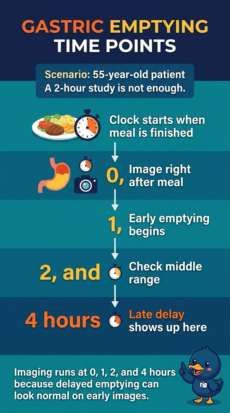
Late emptying is the whole reason this study exists, and it often hides behind normal-looking early images. Your patient's comfort says little about gastric retention, so a smile at hour two doesn't close the book. A car that idles smoothly for two minutes can still refuse to move at the stoplight.
Full Explanation
The 0-, 1-, 2-, and 4-hour schedule is best because gastric retention may appear acceptable on early images yet remain abnormally high at 4 hours; symptom improvement by 2 hours does not establish normal emptying. The schedule ending at 3 hours includes useful serial measurements but substitutes an intermediate endpoint for the standard 4-hour assessment, so it can miss or fail to characterize delayed retention at the validated late time point. The 0-, 2-, and 4-hour schedule retains the decisive late image, but omits the 1-hour measurement used in the recommended standardized series and may provide less information about the emptying trajectory. The 0-, 1-, and 2-hour schedule captures early change, but stopping at 2 hours leaves delayed emptying insufficiently assessed; symptom relief is not a substitute for the late image. Exam caveat: protocols can vary by institution, and results depend on a standardized meal and acquisition method, so distinguish a locally validated protocol from the usual four-hour schedule. Practical check: confirm the patient consumed the specified meal within the required time and that the 4-hour image is acquired and interpreted using the protocol’s criteria.