
A distance runner has three weeks of front-shin pain with normal radiographs. The orthopedist wants assessment of blood flow, the full length of tibial involvement, and imaging limited to the painful leg because cancer has been excluded. Which bone scan plan best fits these three requirements?
Select an answer to reveal the explanation.
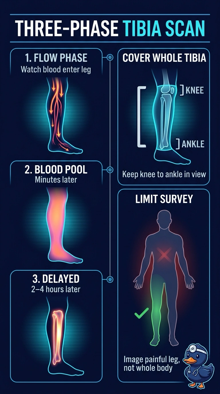
Short Explanation
You want a three-phase study of the painful leg: it checks blood flow and blood-pool activity, then delayed images follow uptake along the whole tibia from knee to ankle, like tracing a trail end to end. Since cancer has been excluded, limiting the scan to that leg avoids an unnecessary whole-body survey; increased uptake can support a stress injury, but it isn’t specific for one.
Full Explanation
A three-phase protocol includes flow images acquired during tracer arrival, blood-pool images acquired shortly afterward, and delayed images that demonstrate osteoblastic activity. Flow and blood-pool findings can assess hyperemia, but they do not by themselves confirm a stress injury. Delayed imaging across the whole tibia shows the distribution of uptake from knee to ankle. Because the question is limited to the painful leg and there is no staging indication, a whole-body survey is not required. A focal three-phase field may miss uptake beyond the area of maximal tenderness, while delayed-only imaging does not assess early perfusion. Increased uptake is sensitive but not specific for a stress injury, so correlate with the clinical findings and other imaging as appropriate. Before the patient leaves the table, confirm that the delayed field includes the entire tibia.