A medical imaging team needs a model that labels every individual pixel of an MRI scan as belonging to a specific tissue type, producing a full-resolution segmentation mask rather than a single image-level label. Which architecture is purpose-built for this pixel-wise task?
Select an answer to reveal the explanation.
Short Explanation and Infographic
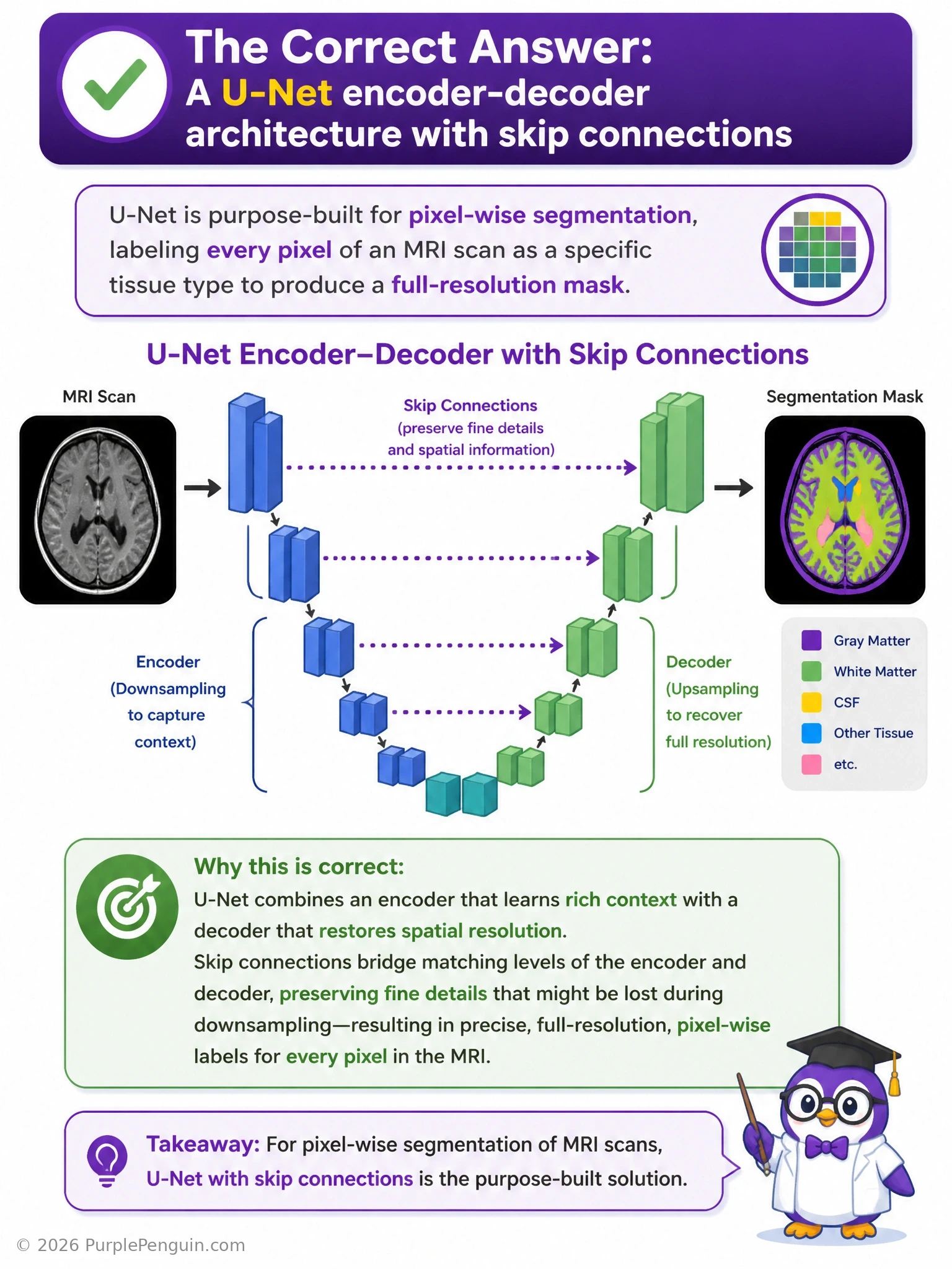
For pixel-by-pixel labeling, you want a U-Net. Picture it as a 'U' shape: the left side (encoder) progressively downsamples the image to capture what's in it, and the right side (decoder) progressively upsamples back to full resolution to say where each thing is, pixel by pixel. The skip connections that hop across the U are the secret sauce — they feed fine spatial detail from the encoder directly into the matching decoder layer, so you don't lose sharp boundaries during all that downsampling and upsampling. A plain classification CNN only gives you one label for the whole image, not per pixel. A GAN without a discriminator isn't really doing adversarial training at all, and it's not built for segmentation. And a single dense layer on raw pixels has no spatial awareness whatsoever — it'll never learn what a tissue boundary looks like.
Full explanation below image
Full Explanation
U-Net is the standard architecture for pixel-wise semantic segmentation, especially in medical imaging. It consists of a contracting path (encoder) that progressively downsamples the input through convolution and pooling layers, extracting increasingly abstract, high-level features while shrinking spatial resolution, followed by an expansive path (decoder) that progressively upsamples back to the original input resolution via transposed convolutions (or upsampling plus convolution). Critically, U-Net includes skip connections that concatenate feature maps from each encoder stage directly to the corresponding decoder stage at the same spatial resolution. These skip connections preserve fine-grained spatial detail (like exact tissue boundaries) that would otherwise be lost during the aggressive downsampling in the encoder, allowing the decoder to produce a precise, full-resolution segmentation mask where every pixel gets its own class label.
The first distractor, a standard classification CNN ending in a single softmax, collapses spatial information into one label for the entire image (e.g., 'this image contains a tumor') rather than labeling each pixel individually — it discards exactly the spatial resolution the task requires. The third distractor, a GAN with no discriminator feedback, is a contradiction in terms and also not the standard tool for this task; GANs (or GAN-inspired segmentation variants) exist, but a plain U-Net with a supervised pixel-wise loss (e.g., cross-entropy or Dice loss) is the standard, purpose-built solution, and removing the discriminator from a GAN just leaves an untrained generator with no meaningful learning signal. The fourth distractor, a single fully connected layer on raw pixels, has no convolutional structure at all and therefore no ability to learn spatial hierarchies or local patterns like edges and textures — it also would require an enormous, impractical number of parameters and would generalize poorly to spatial structure in images.
Memory aid: 'U-Net' literally looks like the letter U when diagrammed — down one side (encode), across the bottom (bottleneck), up the other side (decode), with skip-connection bridges spanning the two sides to preserve detail.