
A 28-year-old motorcyclist arrives on a backboard with a swollen deformed mid-leg after a low-speed crash. The knee and ankle move minimally, distal pulses are intact, and the ED suspects a spiral tibial shaft fracture. The patient cannot stand or rotate the leg freely. Which tibia-fibula projection series should the technologist perform to include both adjacent joints for fracture assessment?
Select an answer to reveal the explanation.
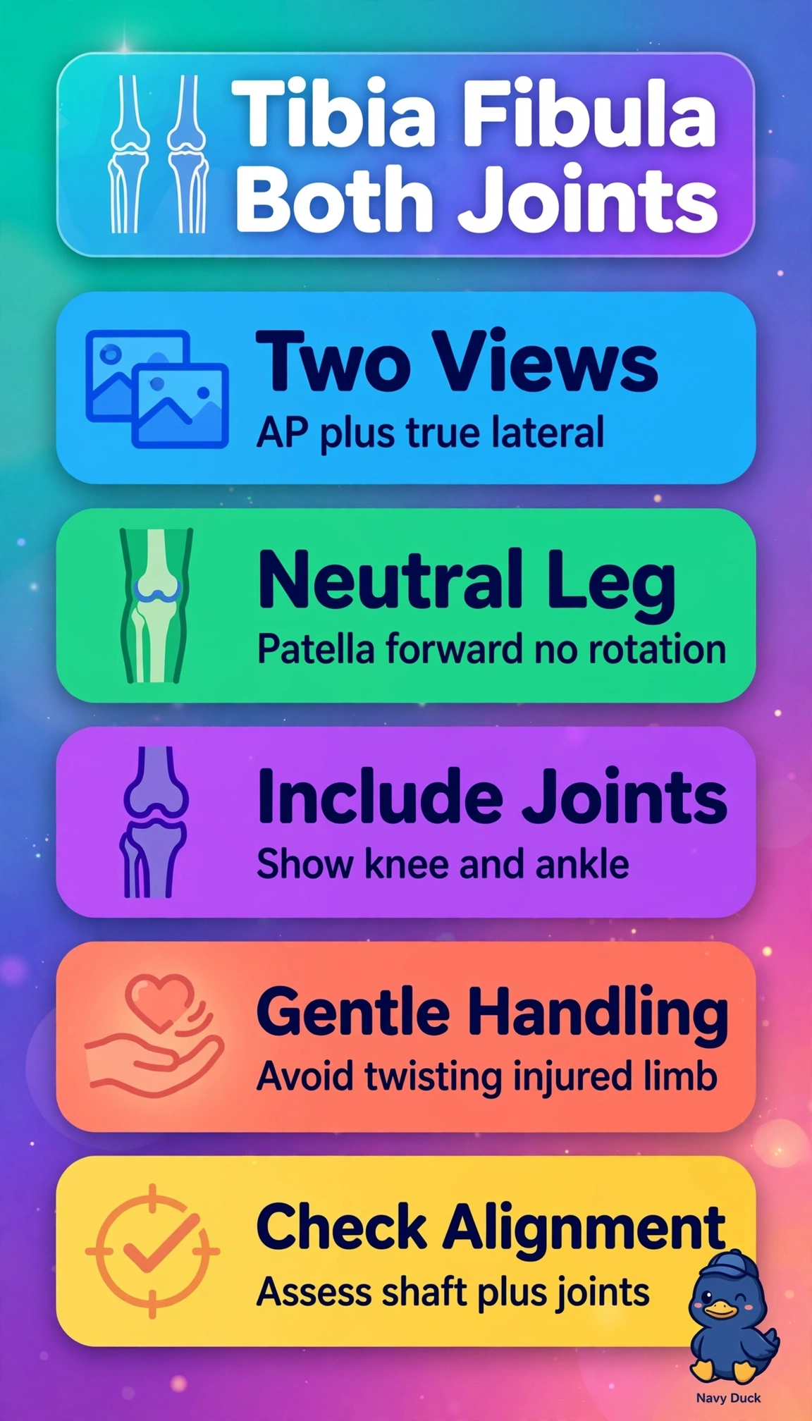
Short Explanation
You need the whole bone plus both ends to catch a sneaky second fracture. Keeping the leg neutral with minimal movement and including the knee and ankle gives you the full span, like checking a whole fence line.
Full Explanation
Perform AP and lateral tibia-fibula views including the knee and ankle, with the leg in neutral rotation and minimal movement. Suspected spiral shaft fracture from torsion requires two orthogonal views of the entire bone to define pattern, displacement and angulation while screening adjacent joints for plateau extension, Maisonneuve fibular neck injury, syndesmotic disruption or pilon involvement. Neutral, minimally handled positioning respects backboard precautions, swelling, deformity, vulnerable neurovascular status and inability to stand or rotate. Weight-bearing AP and lateral views fail because loading an acutely deformed, painful leg in a nonambulatory trauma patient risks further displacement and neurovascular compromise and violates immobilization, although loaded views are routine for chronic alignment or postoperative follow-up. AP and lateral views with 15-degree internal rotation fail because forced rotation for idealized true AP detail requires painful manipulation the patient cannot tolerate and prioritizes ankle profiling over shaft screening. AP and 45-degree oblique views fail because, although an oblique can profile a spiral plane and fibula, it cannot replace a 90-degree lateral to quantify sagittal displacement and guide reduction. An exam caveat is that long-bone trauma requires both adjacent joints on both projections; mortise or isolated-joint logic should raise suspicion even when rotation values sound correct. A practical check is to confirm knee and ankle inclusion on every image, patella forward without forcing rotation, adequate cassette length and neurovascular status after minimal handling; add dedicated joint imaging if effusion, fibular head tenderness or ankle widening appears.