
A 17-year-old with known scoliosis arrives for an erect series and also reports tailbone pain after a fall. The order includes PA erect spine to include C7 to sacrum plus dedicated sacrum and coccyx views. Which dedicated sacrum positioning best demonstrates the sacrum without coccyx overlap?
Select an answer to reveal the explanation.
Short Explanation
You’ve got this—you’re separating erect scoliosis coverage from dedicated sacrum positioning. A cephalad-angle beam projects the pubis away from the sacrum, helping show the sacrum without coccyx overlap.
Full Explanation
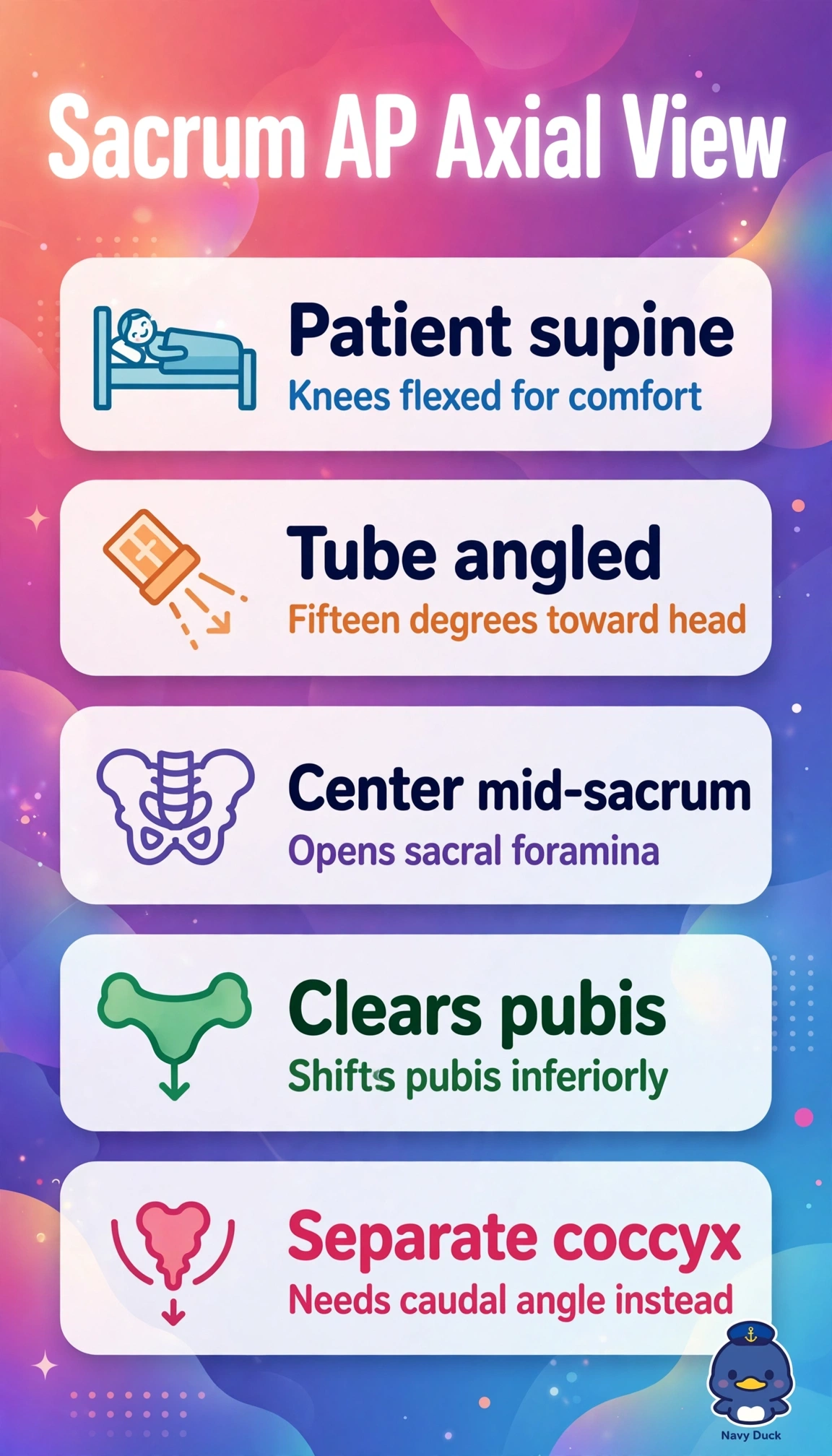
The supine AP axial sacrum view uses a 15-degree cephalad angle to project the pubic symphysis inferiorly, reducing overlap and showing the sacral foramina and segments; this is the dedicated view that best demonstrates the sacrum without coccyx overlap. The scoliosis series has a different purpose: the erect PA spine image includes C7 through the sacrum for spinal assessment, but it does not replace dedicated sacrum positioning. The supine AP axial coccyx view uses a 10-degree caudal angle for the anteriorly tipped coccyx; that angle is intended for coccyx imaging and would not provide the desired sacral projection. A prone PA sacrum image with a perpendicular beam lacks the axial separation needed to move the pubis clear of the sacrum, risking overlap and reduced sacral detail. An oblique SI joint view profiles a sacroiliac joint rather than showing the full sacrum in frontal projection, so it cannot substitute for a dedicated sacrum image. Exam caveat: confirm whether the request is for sacrum or coccyx, since their axial angles differ. As a practical check, center at mid-sacrum and assess whether the sacral foramina are visible and the pubis does not obscure the sacrum.