
A service engineer asks a lead technologist to demonstrate routine compliance for a general radiographic unit used for adult chests, with state inspection approaching and skin dose concerns raised. Which beam restriction and filtration practice should the technologist demonstrate to reduce dose under NCRP 102 principles?
Select an answer to reveal the explanation.
Short Explanation
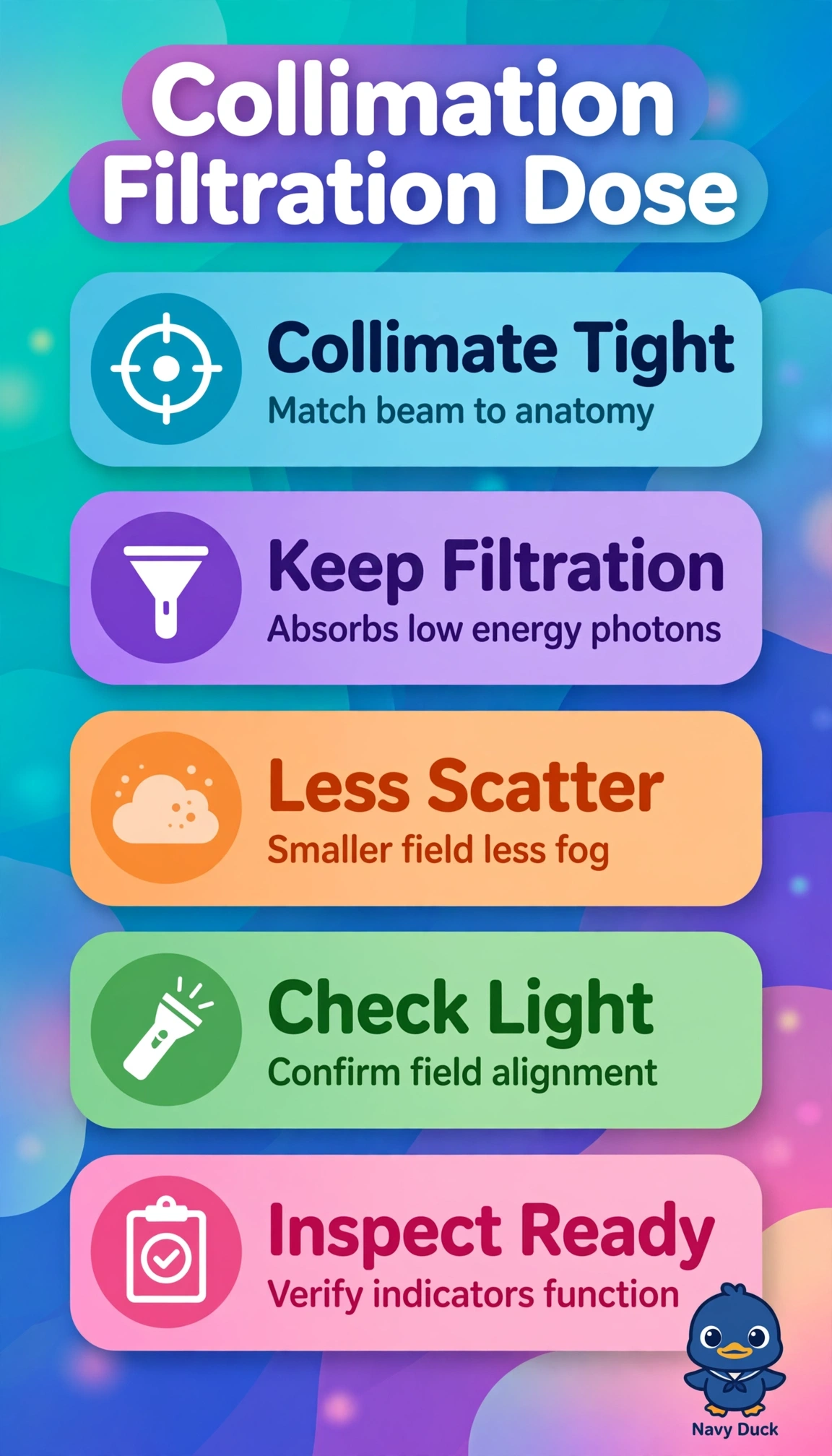
You're trimming beam to what matters. Collimation plus filtration cuts skin dose without hurting the image.
Full Explanation
The correct practice combines two independent NCRP 102 dose controls: tight mechanical collimation limits the primary beam to the anatomy of interest, which directly reduces irradiated skin area, scatter production, and integral dose, while specified aluminum filtration preferentially absorbs low-energy photons that would otherwise contribute only to skin entrance dose without reaching the receptor. The standard 14x17 field choice preserves specified filtration and reflects legitimate concern about cutoff and repeats, but it fails the restriction principle because routinely irradiating full cassette size when a smaller coned field would suffice increases tissue irradiated and scatter despite good filtration. The tight collimation plus copper filtration choice achieves excellent restriction and reflects real low-dose fluoroscopic practice where copper hardens the beam, but it fails the compliance principle because inspection for a general radiographic unit requires manufacturer-specified aluminum filtration and measured half-value layer, and substituting non-specified filtration can alter output, contrast, and technique calibration. The automatic positive beam limitation choice maintains specified filtration and reflects modern room practice, but it fails the active restriction principle because automatic sizing only prevents gross oversizing and does not replace deliberate manual coning to the clinical interest for each adult habitus. An exam caveat is that adequate lung coverage including apices and costophrenic angles is still required, so tight means smallest field that answers the clinical question, not aggressive clipping that forces repeats. A practical check is to demonstrate light-field to x-ray-field congruence, exercise manual shutters to the anatomy, and confirm filtration and half-value layer documentation matches the tube housing chart before inspection.