
An interventional team uses C-arm fluoroscopy to place a peripherally inserted central catheter in a cachectic adult with difficult arm veins, sterile field ready, and tip position must be confirmed. Which fluoroscopic endpoint should the radiographer recognize to confirm correct PICC placement?
Select an answer to reveal the explanation.
Short Explanation
You’re aiming for a central tip, not one that sits too high or extends too deep into the heart. Cavoatrial placement supports reliable dilution while helping you avoid cardiac irritation.
Full Explanation
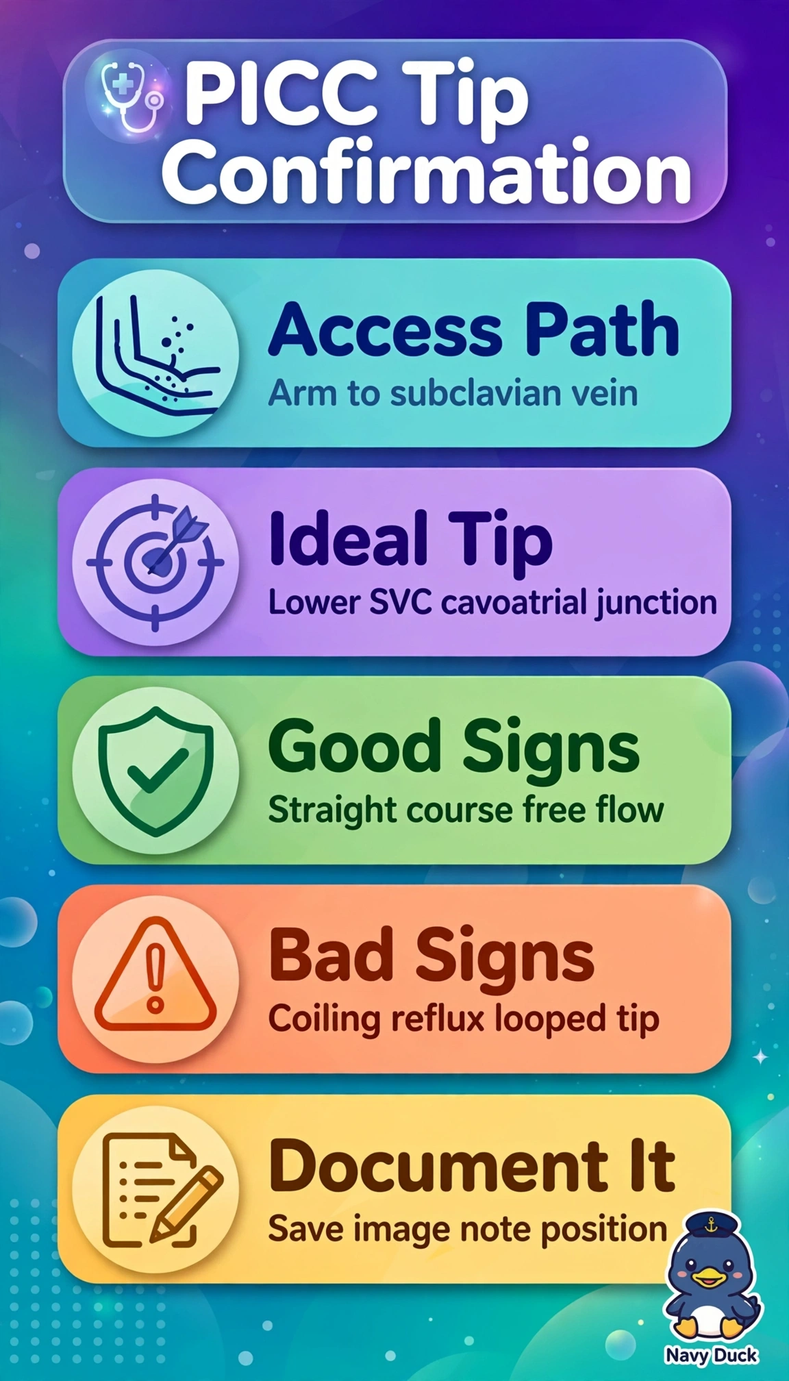
The correct endpoint is the catheter tip in the lower superior vena cava near the cavoatrial junction: this high-flow central position supports immediate hemodilution and stable long-term function, avoids peripheral thrombophlebitis, and keeps the tip outside the heart to reduce arrhythmia and perforation risk. A small contrast injection should show free antegrade flow without coiling, supporting intraluminal placement and a correct course rather than wall abutment or entry into a tributary. A tip in the proximal superior vena cava fails by being too high: despite brisk flow and no coiling or kinking, it is less central, with slower flow, greater thrombosis and malfunction risk, and inadequate dilution for vesicants. A tip in the mid right atrium fails by being too deep: free flow and a straight course do not offset intracardiac risks of arrhythmia, valvular injury, and perforation. A static last-image hold without contrast fails to verify intraluminal position, antegrade clearance, or absence of extravasation, spasm, or sheath effect. Exam caveat: do not apply PICC positioning requirements to dialysis, port, or femoral catheters, which may intentionally use the atrium or inferior vena cava. Practical check: use low-dose live fluoroscopy and a gentle dilute contrast injection during quiet respiration; confirm rapid washout without stasis, reflux, or looping, then secure the catheter sterilely and archive images.