
A technologist compares two lateral elbow images: one crisp with sharp trabeculae using a small focal spot, close receptor, and tight collimation, and one blurry with exaggerated condyles using a large focal spot, 8-inch OID from flexion contracture, and angled beam. Ordering provider questions the difference. Which image quality analysis best separates subject contrast, spatial resolution, and distortion factors?
Select an answer to reveal the explanation.
Short Explanation
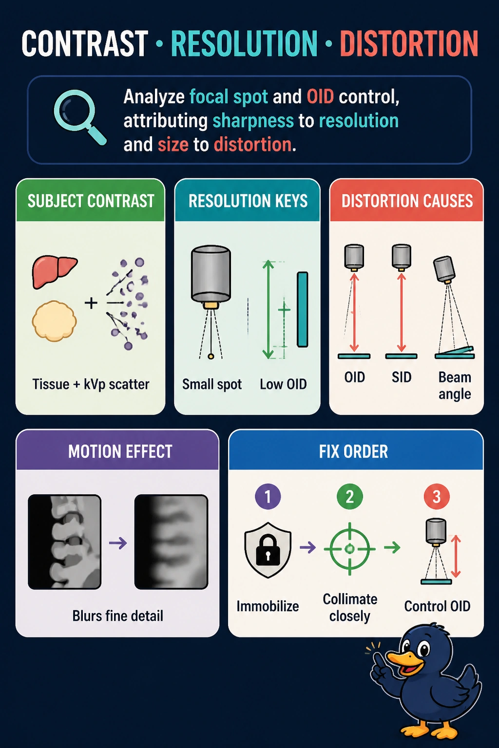
You're sorting sharpness from size from shades of gray. Small spot, small gap keeps edges crisp and anatomy true.
Full Explanation
Spatial resolution is governed by focal spot size, OID, SID, and motion, with small focal spot and minimal OID preserving trabecular detail, distortion encompasses magnification and shape alteration from OID, SID, and beam alignment, so elevated OID with angulation exaggerates condylar size and shape, while subject contrast arises from tissue differences in attenuation influenced by kVp and scatter, not from geometry. The kVp and mAs adjustment approach confuses categories because kVp alters gray-scale contrast and mAs alters receptor exposure and noise, neither of which corrects geometric blur or magnification from large spot and large OID. The SID and grid ratio approach overattributes sharpness to scatter cleanup because grids improve contrast by removing scatter but do not restore resolution lost to geometric unsharpness, and SID alone cannot fully compensate for an 8-inch OID with large spot. The processing window approach mislabels magnification as contrast and blur as distortion because windowing adjusts displayed contrast and brightness after acquisition but cannot undo acquired geometric magnification or unsharpness. Exam caveat is to map tissue and kVp to subject contrast, focal spot OID SID motion to resolution, and OID SID angulation to distortion. Practical check is to use smallest spot, minimize OID, maximize SID within exposure limits, immobilize, and collimate to reduce scatter.