
A clinic repeats a finger radiograph for trabecular detail, but the image shows low subject contrast and blurred margins. The setup used large focal spot, 8-inch OID from a sponge, angled central ray, and high kVp. Which factor set best improves subject contrast, spatial resolution, and shape accuracy together?
Select an answer to reveal the explanation.
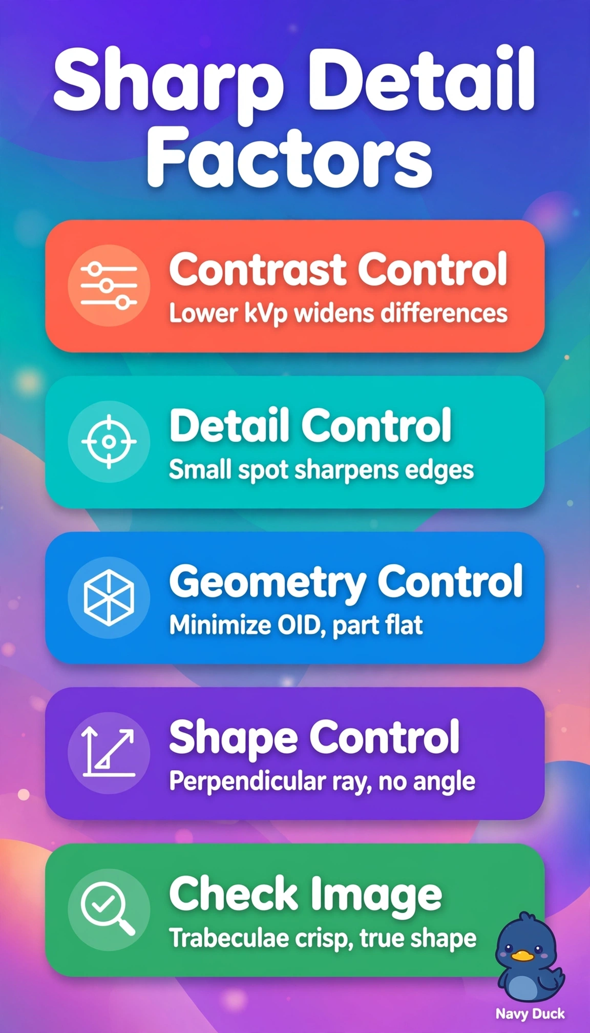
Short Explanation
You’re tightening every link that blurs tiny bone. Lower energy boosts tissue differences while a tight geometry keeps edges true, so resolution comes from small spot and close contact.
Full Explanation
Subject contrast improves with lower kVp because differential attenuation between bone and soft tissue widens, spatial resolution improves with a small focal spot and minimal OID because penumbra and magnification shrink, and shape accuracy improves with a perpendicular central ray and part parallel to the receptor because elongation and foreshortening are minimized. Higher kVp with large spot, increased OID and angled beam degrades all three by lowering differential attenuation while enlarging penumbra and distorting shape, so it fails despite increasing penetration. Higher kVp with small spot but increased SID alone and steep oblique alignment keeps resolution partially but still lowers subject contrast and introduces distortion, so it fails by misapplying SID as a substitute for OID control and angulation. Lower kVp with large spot, increased OID and angled beam preserves some contrast but sacrifices recorded detail and shape, so it fails by retaining blur and distortion despite correct energy. Exam caveat is that ARRT separates subject contrast from image contrast and tests OID versus SID effects distinctly. Practical check is to place the finger flat on the receptor, remove OID, select small spot, lower kVp, and align the beam perpendicular.