
A 78-year-old inpatient fell from bed onto the hip, has pelvic pain and shortening, is hemodynamically stable, can tolerate brief positioning, and the physician suspects pelvic ring disruption needing superior and inferior displacement detail. Which pelvis projection combination best demonstrates pelvic ring integrity and displacement?
Select an answer to reveal the explanation.
Short Explanation
You're mapping the ring from above and below, not just the hip joint. Inlet plus outlet tilts reveal front-to-back and up-and-down shifts with ring-continuity logic like checking a pretzel from two angles.
Full Explanation
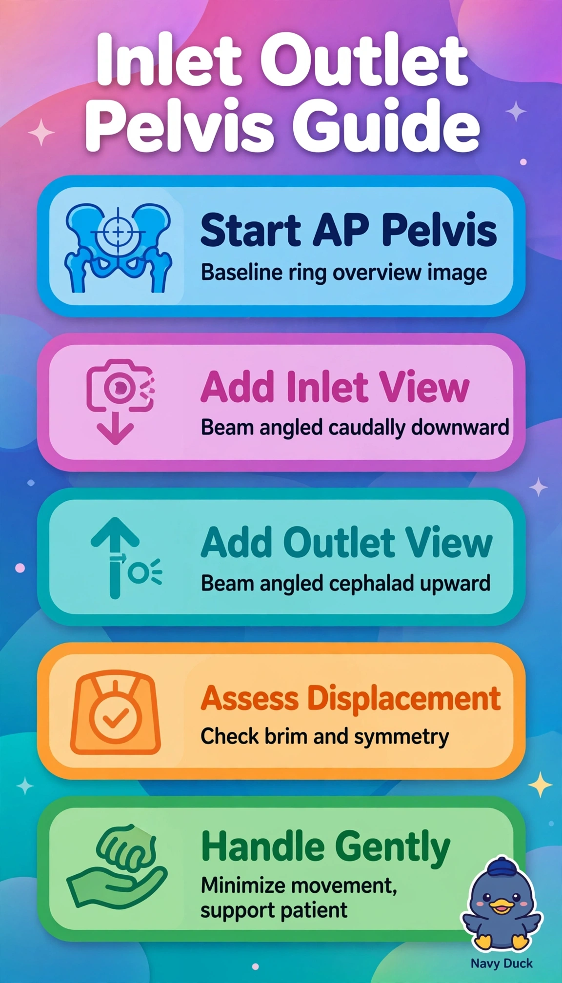
The routine AP pelvis shows symmetric ring, pubic rami, sacroiliac joints and hips, while the supplemental inlet view using caudal angulation profiles the pelvic brim and sacral promontory to demonstrate anterior-posterior displacement and internal rotation, and the outlet view using cephalad angulation opens obturator and sacral foramina to demonstrate superior-inferior translation, vertical shear and sacral impaction, directly meeting the need for superior and inferior detail in a stable patient able to tolerate brief positioning. The Ferguson sacroiliac combination is standard for inflammatory joint disease but isolates the joints with angled centering and lacks whole-ring orthogonal vectors, so vertical displacement can be underestimated. The AP plus inlet-only combination is used for rapid screening but without outlet assessment inferior migration and symphyseal vertical offset remain hidden. The erect weight-bearing inlet-outlet combination is reserved for chronic stability assessment but is contraindicated by acute pain and shortening because loading risks further fracture displacement and hemorrhage. Exam caveat is inlet reflects in-plane anteroposterior shift while outlet reflects vertical shift with shortening signaling vertical shear. Practical check is supine positioning, correct angles (about 40 degrees caudad for the inlet and about 20 to 35 degrees cephalad for male or 30 to 45 degrees for female patients on the outlet), pain reassessment between exposures and CT correlation if needed.