
A critical-care nurse places an enteric feeding tube at bedside and requests radiographic confirmation before feeding, while an interventional team uses mobile fluoroscopy to advance a PICC toward the superior vena cava in another room. Both need imaging guidance. Which radiographic criterion confirms correct placement for enteric tube feeding versus PICC tip position?
Select an answer to reveal the explanation.
Short Explanation
You keep patients safe by knowing where the tip sits. Tip location is everything, like checking a straw reaches the cup before you sip.
Full Explanation
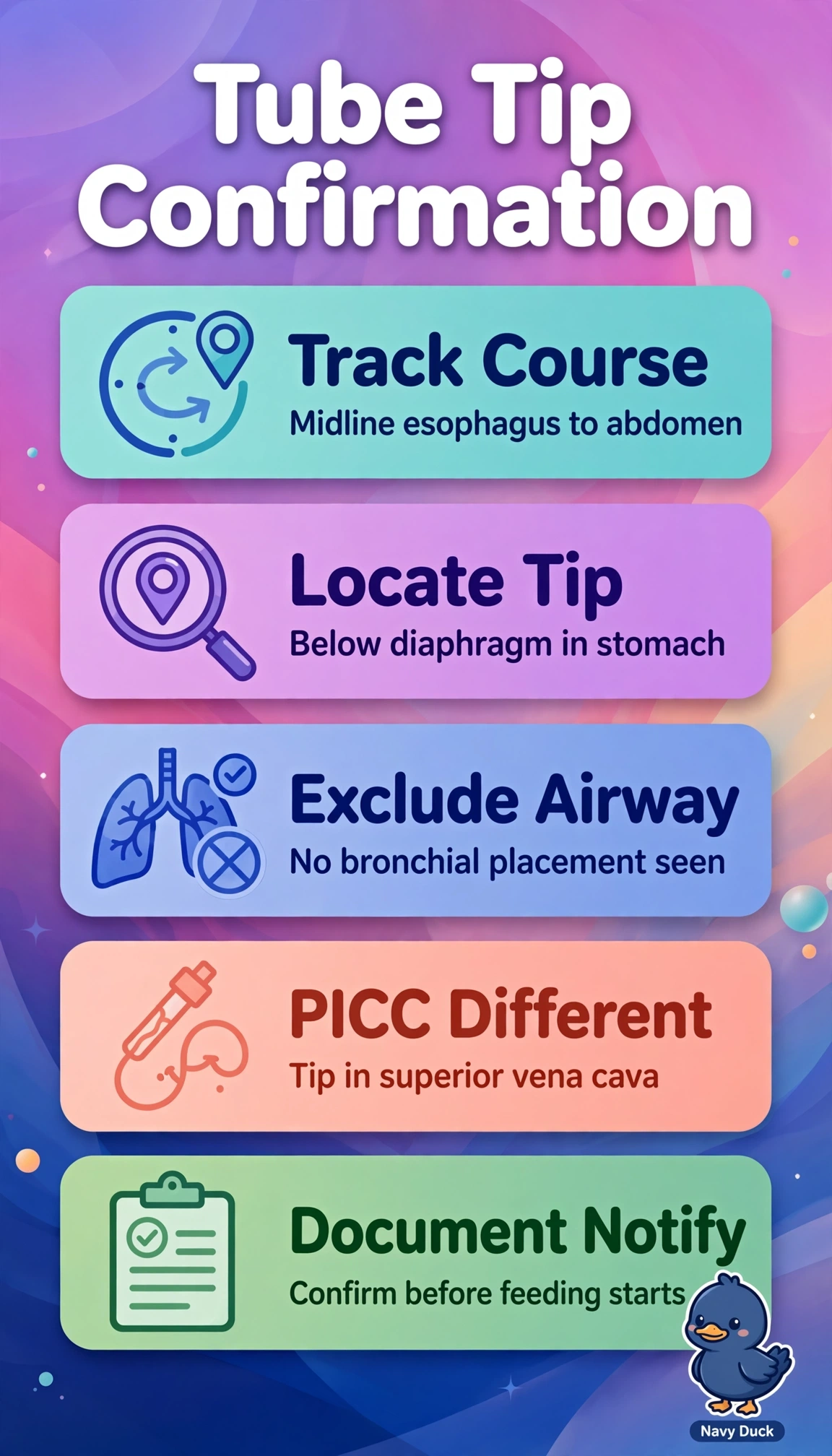
Correct enteric feeding requires radiographic visualization of the tube coursing midline through the gastroesophageal junction with tip clearly below the diaphragm in stomach or duodenum before any feed, which prevents pulmonary or esophageal instillation that bedside tests alone cannot exclude. The criterion describing PICC tip in lower superior vena cava at the cavoatrial junction reflects sound infusion practice for dilution and central access, but it answers the vascular procedure rather than the enteric safety question and cannot authorize feeding. The criterion placing the enteric tip in distal esophagus above the diaphragm fails by level because esophageal position remains above the sphincter with high reflux and aspiration risk despite an otherwise correct course. The criterion accepting auscultatory whoosh for gastric position fails by method because transmitted epigastric sounds occur with bronchial and esophageal malposition and standards require radiography before initial use in high-risk patients. Exam caveat is to separate screening adjuncts from confirmation and vascular from enteric landmarks, remembering diaphragm crossing defines gastrointestinal placement while carina and cavoatrial junction define PICC goals. Practical check is to trace the opaque tube from neck to tip, confirm no coiling in pharynx or bronchus, verify tip below the left hemidiaphragm, and document readiness to feed.