
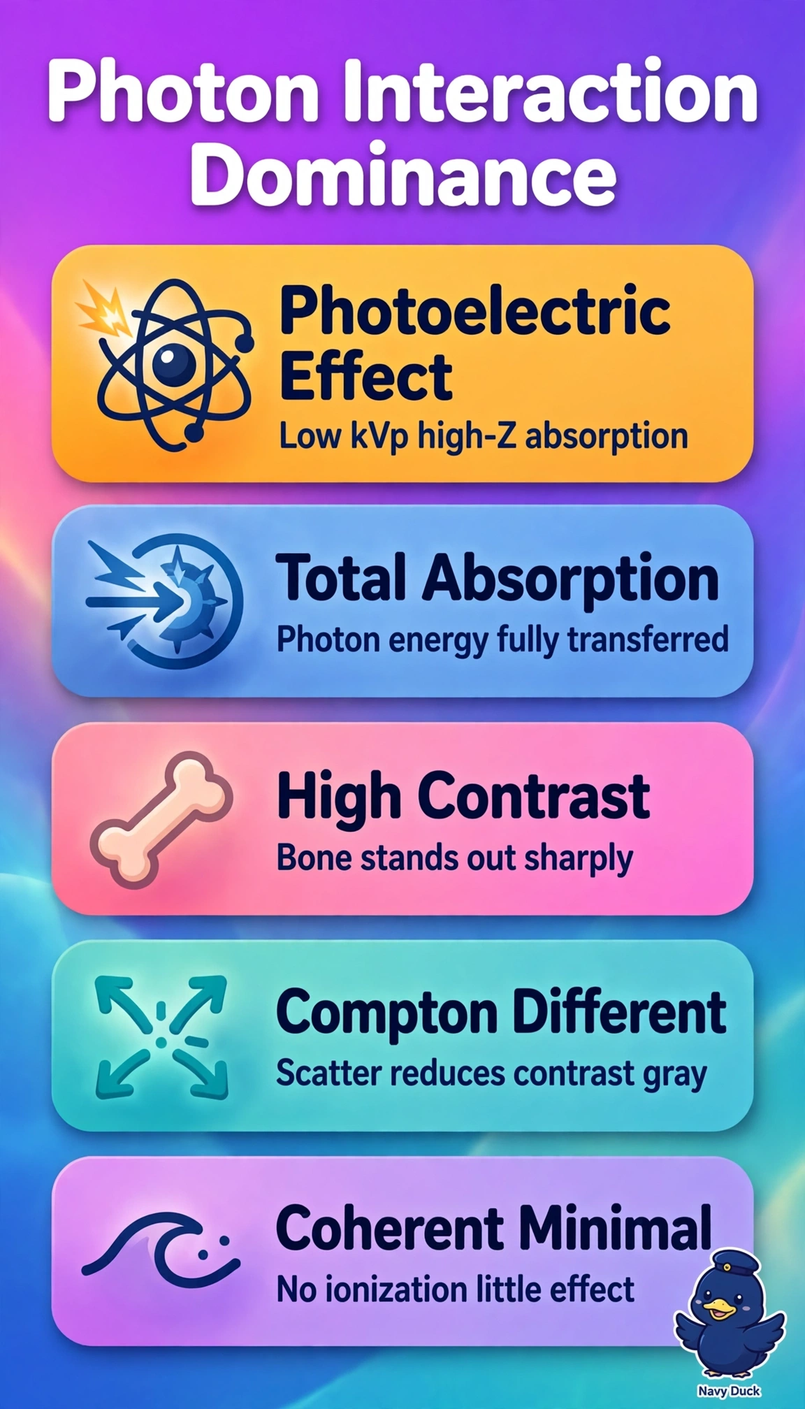
A 22-year-old undergoes a low-kVp hand radiograph with high contrast between bone and soft tissue, while another patient has a high-kVp chest image with more scatter and gray tones. The physicist asks why bone stands out so sharply at low energies. Which photon interaction explains strong bone absorption at low kVp with complete photon absorption?
Select an answer to reveal the explanation.
Short Explanation
You can crack this by linking low kVp to bone brightness. Photoelectric absorption soaks up the whole photon in bone like a sponge, boosting contrast.
Full Explanation
At low kVp photoelectric probability scales approximately as Z cubed over E cubed, so high effective-Z cortical bone with Z near 13.8 absorbs far more completely than low-Z soft tissue, with the entire photon energy transferred to an ejected inner-shell photoelectron and deposited locally to create sharp high-contrast shadows. The Compton-in-bone description fails by interaction physics because Compton involves outer-shell electrons with only partial energy transfer and scattered photon escape, predominates at higher energies and in low-Z tissue, and provides little Z-dependent contrast. The photoelectric-in-soft-tissue description fails by tissue dependence because soft tissue effective Z near 7.4 yields dramatically lower photoelectric cross-section despite identical low kVp and complete absorption, which is why soft tissue remains radiolucent on the hand image. The photoelectric-in-bone-at-high-kVp description fails by energy dependence because at chest energies the photoelectric cross-section collapses and Compton scattering dominates, producing scatter, gray tones, and reduced bone contrast. For exams, remember that completeness of absorption alone does not identify the process, since coherent scatter and characteristic re-emission differ, and always verify technique by checking the kVp on the hand versus chest exposures and confirming expected bone-to-soft-tissue contrast without excessive scatter fog.