
After CT simulation of a pelvic target, the physician draws the target volume on the axial images and specifies that the isocenter should be at its center. Your workstation shows linked axial, sagittal, and coronal views with a movable crosshair. You must document the isocenter in the simulation note before the plan moves to dosimetry. How should you place and verify the isocenter relative to the contoured volume?
Select an answer to reveal the explanation.
Short Explanation
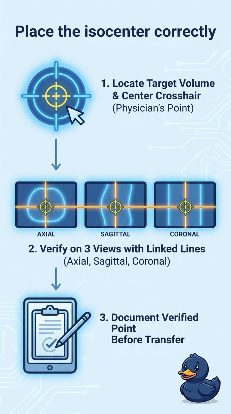
Place your crosshair at the physician’s specified target center, then check that spot in the axial, sagittal, and coronal views before documenting the isocenter. The linked reference lines work like a shared marker, showing you the same location in all three planes.
Full Explanation
The isocenter is the point in space about which the gantry, couch, and collimator rotate. In this scenario, the physician specifies that it should be at the target center, so place the crosshair at that point and verify its position in the axial, sagittal, and coronal views using the linked reference lines. These views help check the point's location in three dimensions, but they do not independently establish the center of an irregular contour; follow the physician's instruction and the simulation workflow. Verifying only the axial view can miss a positional error in another plane. Bony landmarks may not coincide with the specified target center, and skin markers indicate an external position rather than the specified internal target point. Document the verified coordinates before the plan moves to dosimetry, according to local workflow.