
A liver SBRT patient has target motion larger than the 5 mm setup margin, breath-holds run out before the beam time ends, and the abdominal compression plate is painful over the treatment site. Respiratory gating with an external breathing surrogate is available. Which motion-management method best matches this patient's stated limits?
Select an answer to reveal the explanation.
Short Explanation
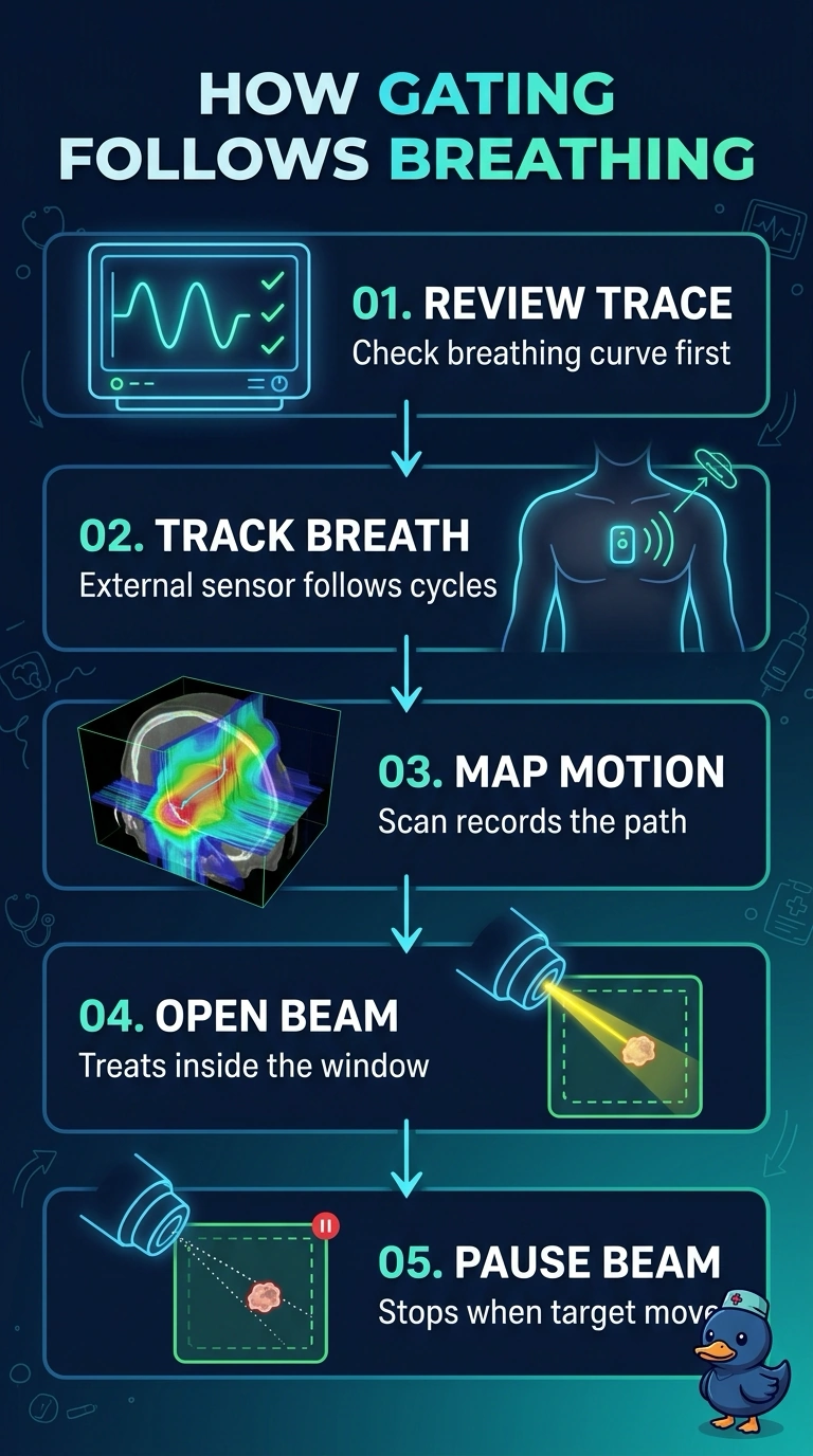
Gating is the only choice that fits how this patient actually breathes, since it treats only the quiet part of the breathing cycle. The stated limits quietly close the other doors: the plate hurts, the holds run out, and the margin's already too small. You still get motion control without asking for anything painful.
Full Explanation
Respiratory gating with the available external breathing surrogate best addresses the central constraint: it permits treatment during free breathing while restricting beam-on time to the respiratory window in which the lesion remains within the allowed motion. It avoids relying on the inadequate 5 mm setup margin to cover the full excursion, does not require the painful compression plate, and does not depend on sustaining a breath-hold throughout beam delivery. A customized, broad-contact compression setup is a recognized way to reduce liver motion and image guidance can support positioning, but it still applies pressure through the painful treatment-site region; refining the plate does not resolve the stated intolerance. Phase-specific treatment at end expiration can be reproducible, yet a 5 mm expansion around that phase does not encompass the larger excursion across respiration, so it risks geographic miss if the target leaves the selected phase during delivery. Breaking delivery into short breath-hold segments with recovery pauses is also established practice, but the stated holds end before the required beam time is complete; pauses do not make the patient able to sustain the necessary holds. Exam caveat: gating is appropriate only when the surrogate reliably correlates with internal tumor position and the motion window is adequately defined. Practical check: verify that the external signal tracks the lesion on simulation imaging and confirm the gating window and beam interlock before treatment.