
Before a first fraction, a therapist reviews a new chart holding a history and physical describing a right breast mass with a suspicious axillary node, a mammography report noting a 2.8 cm spiculated mass, a bone scan showing one sclerotic rib lesion, and a core biopsy pathology report reading invasive ductal carcinoma. Which document should confirm the histology listed on the treatment record?
Select an answer to reveal the explanation.
Short Explanation
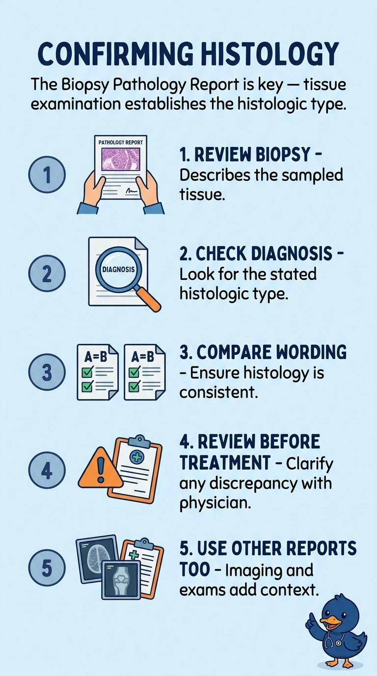
Your source for histology is the core biopsy pathology report, which examines the tumor tissue—like checking what’s inside rather than judging the package. Imaging shows the tumor’s location and extent, while the history and physical summarizes the clinician’s assessment; if the wording differs, you should clarify with the responsible physician.
Full Explanation
The core biopsy pathology report should confirm the histology on the treatment record because it examines sampled tumor tissue and identifies the cancer’s histologic type; here, it documents invasive ductal carcinoma. The mammography report is an imaging-based assessment that establishes the tumor’s site and size—such as the 2.8 cm spiculated mass—for planning, but imaging appearance does not establish histology. The history and physical is a clinical-summary approach: it records the physician’s diagnosis and disease status, but it is not the tissue-based source for confirming histologic type. The bone scan is a distant-disease assessment approach; it documents a finding relevant to evaluating possible spread, such as the sclerotic rib lesion, rather than defining the breast tumor’s histology. Exam caveat: these records provide complementary information, so a suspicious imaging finding or clinical diagnosis should not be treated as a substitute for tissue confirmation. Practical check: compare the treatment record’s histology with the biopsy pathology wording, and if they differ, clarify the discrepancy with the responsible physician before proceeding.