
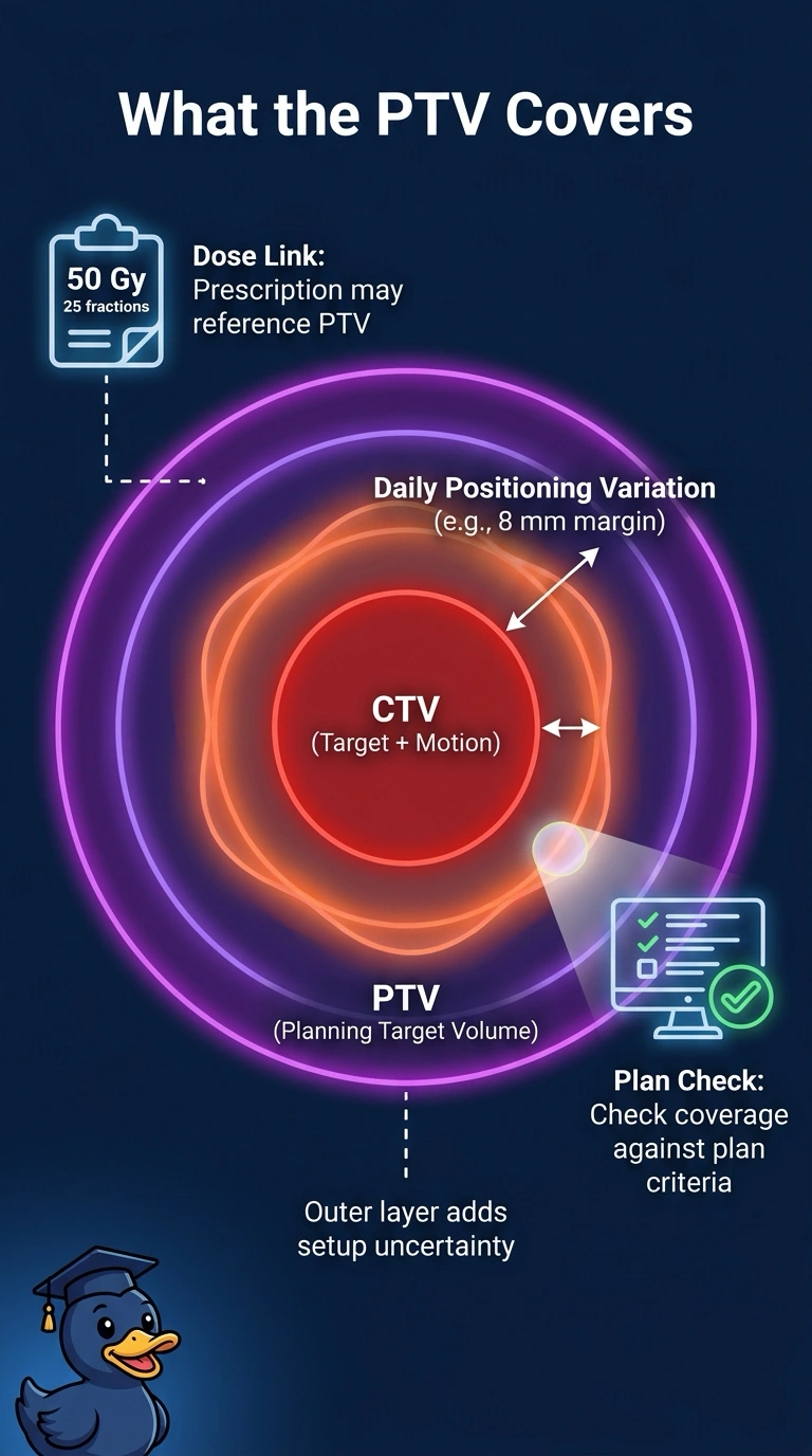
On the planning CT, the team contoured the visible mass, expanded it 8 mm for possible subclinical disease, added the full breathing motion envelope, and then added setup uncertainty. The prescription delivers 50 Gy in 25 fractions to this final contour. Which target volume does the final contour represent?
Select an answer to reveal the explanation.
Short Explanation
Those contours nest inside each other, and each layer absorbs a different kind of uncertainty. Adding setup uncertainty at the end is what makes that outer layer a PTV. It's the last shield in the stack: everything inside it, plus your daily wobble.
Full Explanation
Each target volume is defined by the kind of uncertainty it is meant to absorb, and your final step added setup uncertainty, which is characteristic of a planning target volume. Stopping at the gross tumor volume would limit the contour to what imaging shows and ignore the ring of suspected subclinical disease. Building only the clinical target volume captures that microscopic extension but still leaves respiratory motion and daily placement error outside the boundary. Using the internal target volume as the final contour covers the breathing envelope yet omits uncertainty in how the patient is positioned each day, so coverage would be judged against a narrower edge than the prescription intends. Exam caveat: when motion is represented with an ITV, the PTV is built from that ITV plus a setup margin. If motion is not explicitly represented with an ITV, appropriate margins must account for it as well as setup uncertainty. Practical check: open the structure definitions, confirm which contour the prescription line references, and verify the dose is reported to that exact volume.