
A patient with a lower-lobe lung tumor is booked for simulation. The tumor moves noticeably with each breath, and the patient struggles to follow coached breath-hold instructions. CT simulation with breathing-motion imaging is available, while these machines cannot deliver respiratory gating. Which simulation, positioning and immobilization approach best accounts for the target's motion?
Select an answer to reveal the explanation.
Short Explanation
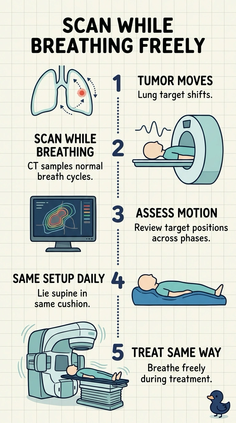
You’ll want a breathing-motion CT while you breathe freely, then treat supine with the same free-breathing approach. A breathing-motion CT is like a long-exposure photo of a moving car: it captures the target across its path, not just at one instant.
Full Explanation
The appropriate approach captures the tumor’s respiratory excursion on a breathing-motion CT and uses a consistent supine, free-breathing setup for treatment. This suits a patient who has difficulty following coached breath-hold instructions and does not depend on respiratory gating, which the treatment machines cannot provide. Applying abdominal compression only during treatment introduces a motion-reduction technique after simulation; the anatomy and tumor motion represented by the scan may therefore differ from those at delivery. Planning from the end-expiration phase alone while treating freely uses one phase to define the target but delivers through the full breathing cycle. Without gating or a target that encompasses motion, the tumor can move outside the planned volume. Aligning daily to the tumor’s average position and using a tight margin also fails to cover its full excursion; alignment cannot substitute for motion coverage. Exam caveat: a 4D CT records motion but does not itself guarantee that the plan covers the full motion envelope or that treatment reproduces the simulation setup. Practical check: review the motion dataset and confirm that the planned target and margins encompass the observed excursion under the same positioning and breathing conditions used for treatment.