
A 4-year-old with fever and known vesicoureteral reflux is referred for renal cortical imaging to evaluate scarring and differential function. The child will be awake and must stay still for a single study. Which tracer, timing, and views should be used for this renal cortical study?
Select an answer to reveal the explanation.
Short Explanation
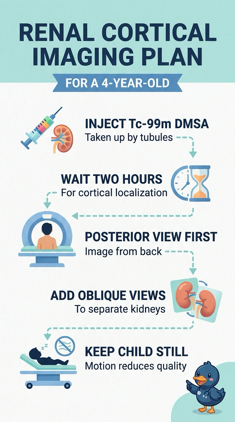
Use Tc-99m DMSA, then wait about two hours before taking posterior and oblique images; you’ll need to stay still for this single study. DMSA works like a highlighter for the kidney’s cortex, helping show scars and each kidney’s share of function.
Full Explanation
Tc-99m DMSA is taken up by proximal tubular cells in the renal cortex and retained for hours, so imaging roughly two to three hours after injection generally provides good cortex-to-background contrast and can reveal photopenic areas associated with scarring. Posterior and oblique views help assess the kidneys and separate them from overlapping structures. A dynamic MAG3 acquisition measures transit and drainage through the collecting system, so it answers an excretion question rather than a cortical one. Imaging DMSA at thirty minutes may leave more blood-pool activity in the background, making the cortex less distinct even though uptake has begun. A diuretic renogram with DTPA evaluates washout and split function during a drainage study, but it does not provide the stable cortical retention needed for scar detection. On the exam, the words cortical, scarring, or differential function should steer you to a cortical agent. Confirm the tracer, timing, and view set on the department protocol before imaging.