
A 4-year-old with a history of febrile urinary tract infection is referred for Tc-99m DMSA renal cortical imaging to look for scarring. The department can schedule imaging 1 hour or 2 to 3 hours after injection and offers planar alone or planar plus SPECT. Which plan is most sensitive for finding a focal cortical defect in this child?
Select an answer to reveal the explanation.
Short Explanation
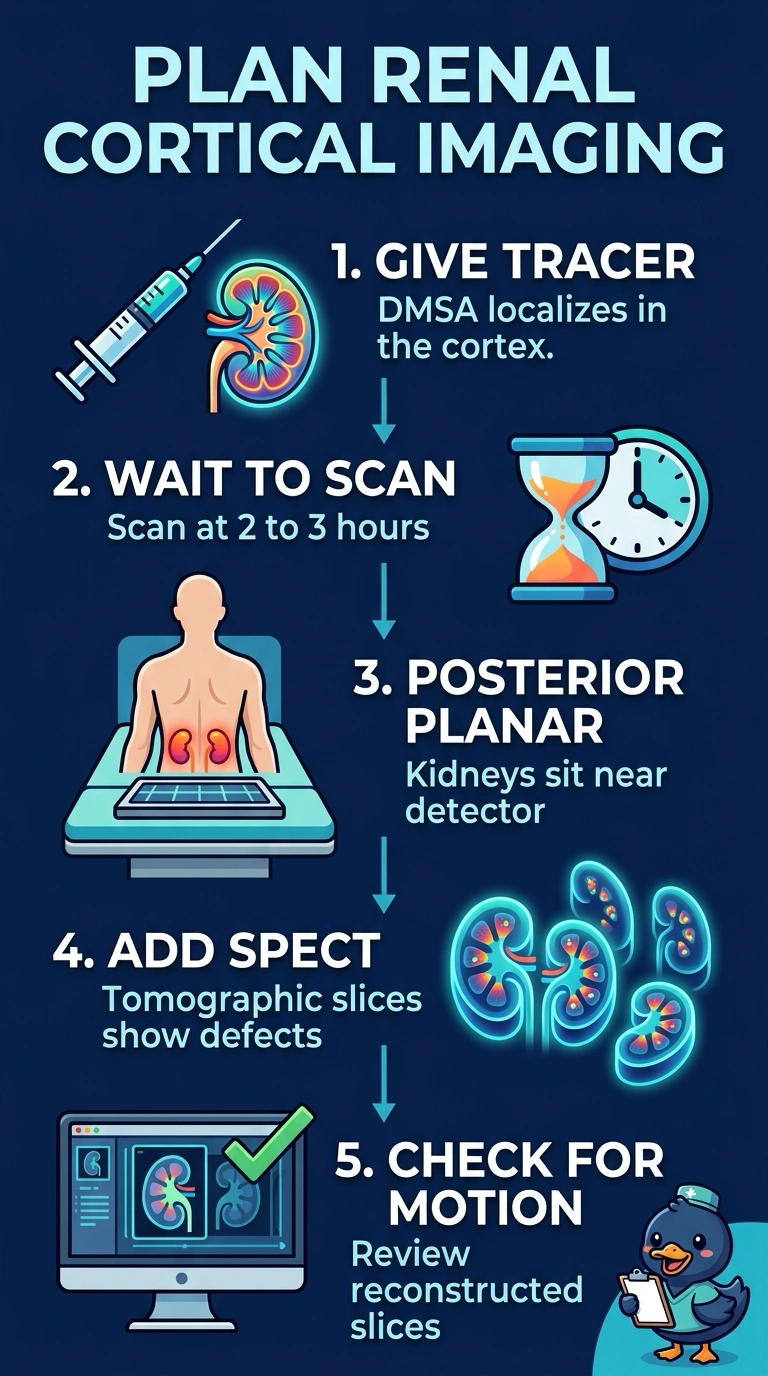
You'll generally improve detection by allowing time for cortical uptake, then adding SPECT rather than relying on a single planar view. Tomographic slices can help reveal defects that overlap normal cortex in a projection image. The 2 to 3 hour window plus SPECT is the most sensitive option here.
Full Explanation
Tc-99m DMSA localizes in the renal cortex, and imaging is typically performed about two to three hours after injection. Posterior planar views are standard, but SPECT can add tomographic information and improve detection of focal defects that may be obscured in projection images. Planar-only imaging at the recommended time can show cortical defects, but it does not provide the additional tomographic information of SPECT. Imaging at one hour is earlier than the usual imaging window and may provide less favorable cortical-to-background contrast. Dynamic acquisition over the first twenty minutes is not the usual protocol for assessing cortical scarring; it is used in other renography protocols to assess perfusion and transit. A young child may not stay still for a long acquisition, so review motion on the reconstructed slices before accepting the study. Confirm the injection time and planned acquisition window on the worklist.