
A child is referred for Tc-99m DMSA imaging after a febrile urinary tract infection, and the history suggests scarring in one kidney. The protocol allows several hours for tracer uptake, and the study must show focal defects and relative function in both kidneys. Which imaging plan best demonstrates renal cortical detail?
Select an answer to reveal the explanation.
Short Explanation
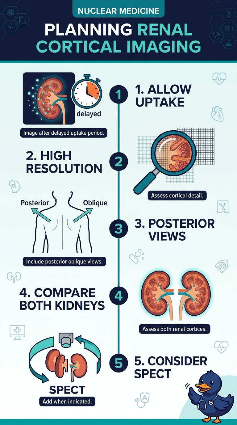
After the tracer has had a few hours to build up, you’ll get the best cortical detail with high-resolution posterior and posterior oblique views of both kidneys, adding SPECT if needed. It’s like comparing both sides of a pair: imaging both kidneys shows focal defects and relative function, while imaging just one can’t give you that comparison.
Full Explanation
The correct plan uses delayed imaging, allowing Tc-99m DMSA to accumulate in functioning renal cortex, and high-resolution posterior plus posterior oblique views to display cortical contours and focal defects while imaging both kidneys. Relative cortical function can be compared between the kidneys; SPECT may be added when planar findings are equivocal or more localization is needed. The anterior-only plan is a recognized projection, but as the sole view it is less suitable for this question: attenuation and overlap can limit cortical detail, and it omits the posterior and oblique projections used to characterize scars. Early dynamic planar imaging is a standard approach for assessing tracer transit and perfusion with appropriate renography agents, but it is mistimed for DMSA cortical imaging; before several hours of uptake, it cannot reliably demonstrate the requested cortical defects or relative cortical function. Imaging only the suspected kidney may help characterize a known lesion, but it cannot provide the required bilateral comparison or relative function. Exam caveat: DMSA protocols and whether SPECT is added vary with the clinical question and local practice; SPECT is not mandatory in every study. Practical check: confirm delayed uptake time, bilateral coverage, adequate posterior/oblique positioning, and that image quality permits comparison before ending acquisition.