
An adult with a known lung tumor reports new pain between the shoulder blades, and the nuclear medicine team must survey the entire skeleton and pinpoint which part of the thoracic spine is taking up tracer. Which bone imaging plan best meets both needs?
Select an answer to reveal the explanation.
Short Explanation
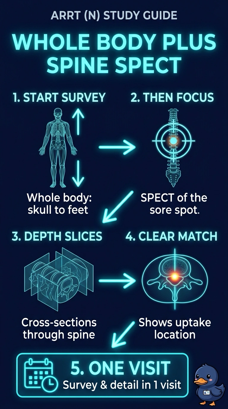
You need whole-body images to check whether disease is anywhere in the skeleton, then targeted SPECT to help pinpoint uptake within the thoracic spine. Together, they give you broad coverage and cross-sectional localization in one visit—a survey jacket with a built-in magnifier. Three-phase imaging, a limited study of the painful area, or planar spot images would leave your metastatic survey or localization unfinished.
Full Explanation
Whole-body images from skull to feet survey the entire skeleton, which is important when an adult with a known lung tumor needs evaluation for spread to bone. Targeted SPECT through the symptomatic thoracic spine then provides cross-sectional slices that can help localize uptake to the vertebral body or posterior elements, which may overlap on planar images. Together, these images meet both the survey and localization needs. Three-phase bone scintigraphy of the thoracic spine includes flow, blood-pool and delayed images and can help assess a localized concern such as infection, but it does not survey the entire skeleton. A limited planar and SPECT study of only the painful mid-back, even with the opposite side for comparison, focuses on a regional site and leaves the whole-skeleton survey undone. Whole-body images followed by planar spot images do provide the survey, but planar superimposition cannot reliably assign uptake to a particular part of a vertebra, so the localization request remains unmet. Exam caveat: planar images can show uptake without resolving which vertebral structure is involved. Practical check: confirm that the referral asks for both a whole-skeleton survey and localization at a specific site before choosing the protocol.