
A patient with COPD is being evaluated for lung resection, so the surgeon needs each lung's share of blood flow. The study must also let ventilation be compared with perfusion in the same body positions. Which acquisition plan meets both the matching-view comparison and the quantitative lung percentages?
Select an answer to reveal the explanation.
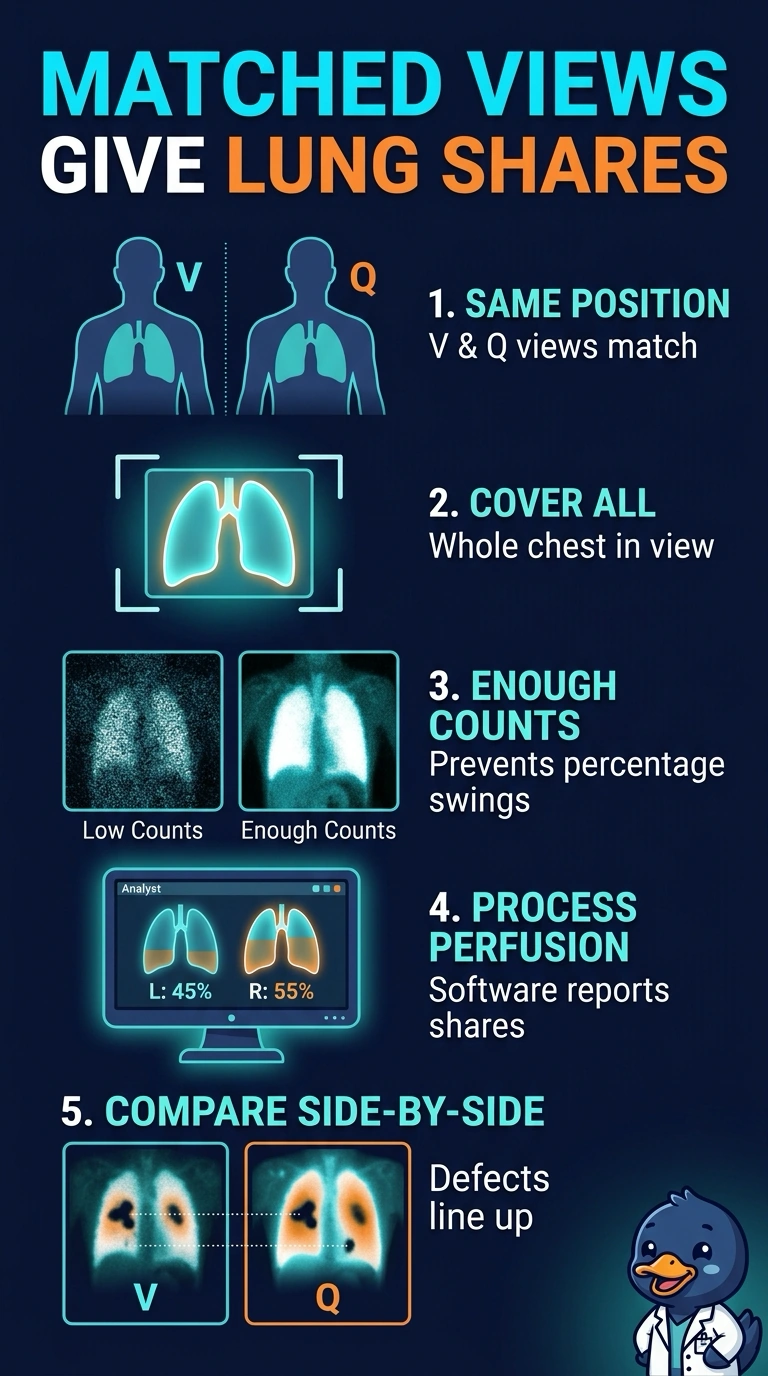
Short Explanation
You've got two jobs here: views that line up and numbers you can trust. Matching positions lets each perfusion defect be compared against the same ventilation image, and sufficient counts keep the quantitative output stable. Skip either piece and the surgeon gets only half of what was ordered.
Full Explanation
A quantitative lung study has to deliver two results at once, and planar acquisition supports both only when geometry and statistics are handled together. Identical projections for ventilation and perfusion place the two data sets in the same frame of reference, so a wedge-shaped perfusion defect can be matched against the corresponding ventilation image rather than compared across different angles. Collecting enough counts in each projection gives the region-of-interest software a stable ratio, because percentages drawn from thin, noisy images swing with statistical variation and can shift a surgical estimate. Acquiring only anterior and posterior perfusion images keeps count statistics strong but removes the ventilation partner, so the study can quantify function yet cannot be read as a matched comparison. Taking matched views with a deliberately short time per view protects the schedule but starves the quantitative step of the counts it needs for reproducibility. Caveat: count targets and software packages vary by manufacturer, so use the criteria stated for your own system. Practical check: before the patient leaves, confirm every perfusion view has a ventilation partner and that the counting statistics look even across both lung fields.