
A 74-year-old with HFpEF and concentric LV wall thickening is referred for Tc-99m PYP imaging to evaluate suspected ATTR cardiac amyloidosis. Serum and urine monoclonal studies are negative and kidney function is stable. The laboratory's protocol starts with a 1-hour acquisition. Which acquisition plan best separates true myocardial retention from residual blood-pool activity?
Select an answer to reveal the explanation.
Short Explanation
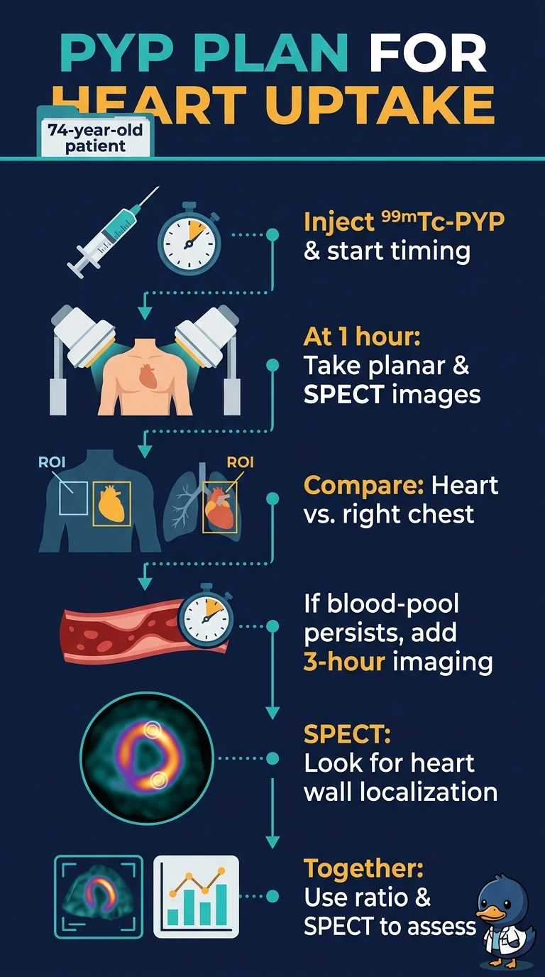
You’ll acquire anterior planar and SPECT images at 1 hour; SPECT helps show whether tracer is in the heart muscle or still in the blood pool. If blood-pool activity persists, add 3-hour imaging, and use the heart-to-contralateral ratio as an adjunct, not proof of myocardial uptake.
Full Explanation
The recommended plan combines anterior planar imaging and SPECT at 1 hour, with 3-hour imaging added if blood-pool activity persists. SPECT localizes apparent cardiac activity to the myocardium or ventricular cavity, while the heart-to-contralateral (H/CL) ratio provides a quantitative adjunct rather than a substitute for image interpretation. A plan using 1-hour planar images and the H/CL ratio but no SPECT cannot reliably separate myocardial retention from blood pool, because activity can overlap the cardiac silhouette. A plan that begins with 3-hour planar and SPECT imaging may help assess slow blood-pool clearance, but omits the requested 1-hour acquisition and initial-plus-conditional-delayed sequence. A plan that includes 1-hour SPECT and conditional 3-hour imaging but treats H/CL as definitive overstates the ratio: acquisition and processing factors can affect it, and it should not override SPECT evidence of blood-pool activity or myocardial localization. Exam caveat: interpret PYP uptake alongside monoclonal-protein testing; a positive scan alone does not establish ATTR when a monoclonal gammopathy is present. Practical check: review SPECT or SPECT/CT slices in multiple planes to confirm uptake in the myocardial walls rather than the ventricular cavity.